Материал: ABC-of-Rheumatology-ABC-Series-

Low Back Pain |
27 |
|
|
|
|
Acknowledgements
Table 4.1 and Box 4.3, and Figures 4.1, 4.2, 4.3, 4.4 and 4.6 are taken from Imboden et al., 2007, with permission. Figure 4.5 is
taken from Standards in Rheumatology: a Suggested Management Plan for Some Common Conditions in Rheumatology. The Medicine Group, 1987.

CHAPTER 5
Pain in the Hip
Andrew J Hamer1 and Jeffrey N Katz2
1Northern General Hospital, Sheffield, UK
2Case Western Reserve University, Cleveland, USA
OVERVIEW
•Osteoarthritis of the hip is common in adults, and osteoporotic hip fractures are epidemic in the elderly.
•Always examine the hip in patients presenting with knee pain, as referred pain from the hip is common.
•Childhood hip conditions require prompt treatment to reduce the risk of problems in later life.
Box 5.1 Important causes of childhood hip pain
•Congenital dislocation of the hip
•Perthes’ disease
•Slipped upper femoral epiphysis
•Septic arthritis
•Transient synovitis or “irritable hip”
•Other arthritides
Hip pain in children
A child with hip disease may not present with pain or a history of trauma but with an unexplained limp. Unexplained thigh or knee pain should also raise the suspicion of hip abnormality. See Box 5.1 for a summary of important causes of childhood hip pain.
Congenital dislocation of the hip
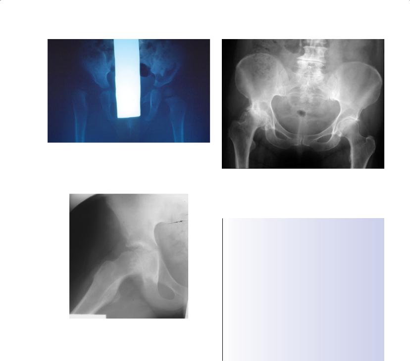
Physical examination and/or ultrasound screening should detect at-risk cases (Figure 5.1), but missed cases may present as a delay in walking, a limp or discrepancy in leg length. Children usually present before 5 years of age. Missed cases may lead to a noncongruent joint and early osteoarthritic degeneration in adulthood.
Perthes’ disease
Perthes’ disease—disintegration of the femoral head, with subsequent healing and deformity of the hip—usually occurs in boys aged 5–10 years. The precise cause is unclear, but segmental avascular necrosis of the femoral head is probably responsible. A limp, hip pain or knee pain may result. Treatment aims to contain the femoral head in the acetabulum to reduce the risks of future osteoarthritis.
Slipped upper femoral epiphysis
This condition is typically seen in overweight, hypogonadal boys, who often present with pain referred to the knee, although girls
ABC of Rheumatology, 4th edn. Edited by Ade Adebajo. ©2010 Blackwell Publishing Ltd. 9781405170680.
may also experience this condition. The diagnosis may be difficult, but a “frog lateral” X-ray radiograph will show the deformity (Figure 5.2).
Surgical stabilization is needed as a matter of urgency to prevent further slippage of the epiphysis. The contralateral hip is at high risk of slippage, and patients and parents should be warned to return if any knee or hip pain occurs.
Septic arthritis
This is relatively uncommon, but it should be suspected in a child who is ill, toxic and unable to walk. Movement of the affected joint is not possible because of pain. Diagnosis is confirmed by raised white cell count and erythrocyte sedimentation rate and perhaps by effusion on ultrasound images. No test is perfectly sensitive or specific, so expert clinical judgement is required. Urgent surgical drainage is vital to reduce the risk of late osteoarthritis. Diagnosis may be particularly difficult in neonates. Staphylococcus aureus is the usual infective organism.
Transient synovitis or “irritable hip”
A reactive effusion may occur in the hip in association with a systemic viral illness. Affected children are not acutely ill and can move the hip, but with some degree of stiffness. An effusion may be seen on ultrasound images and the condition is usually selflimiting and responsive to non-steroidal anti-inflammatory drugs. Distinguishing this condition from septic arthritis can be a challenge, and occasionally these children must undergo aspiration to exclude a septic hip. Perthes’ disease may present in the early stages with an effusion without changes visible on X-ray examination.
28
Pain in the Hip |
29 |
|
|
|
|
Figure 5.1 Anteroposterior radiograph of child with dislocated right hip. Note the lateral displacement of the femur and the poorly developed ossific nucleus of the hip
Figure 5.2 X-ray radiograph of a child’s right hip. Displacement of the epiphysis relative to the femoral neck is easily seen
Other arthritides
Juvenile chronic arthritis may present with hip pain. General management of the arthritic process is important, with physiotherapy to prevent joint contracture. Systemic therapy with disease-modi- fying agents (such as methotrexate, tumour necrosis inhibitor agents) can be very effective. These therapies have important potential toxicities and must be prescribed knowledgeably.
Hip pain in adults
Pain from the hip is usually felt in the groin or lateral or anterior thigh. Hip pain may also be referred to the knee; this may confuse the unwary! Although buttock pain may originate from the hip, the lumbar spine is the usual source. Hip disorders often produce a limp, a reduction in the distance that can be walked, and stiffness.
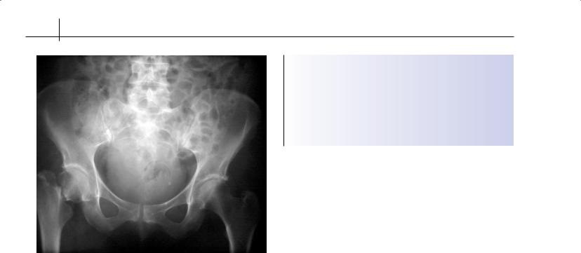
Figure 5.3 Osteoarthritis of the right hip, with joint space loss, subarticular cysts, peripheral osteophytes and subchondral sclerosis
Box 5.2 Causes of hip pain in adults
•Osteoarthritis
•Other arthritides
Rheumatoid arthritis
Psoriatic arthritis
Ankylosing spondylitis
•Hip fracture
•Paget’s disease
•Avascular necrosis
•Malignancy
•Infection
•Painful soft-tissue conditions around the hip
Trochanteric bursitis
Iliopsoas bursitis
Ischial bursitis
Meralgia paraesthetica
Snapping iliopsoas tendon
Torn acetabular labrum
These functional limitations may prevent activities of daily living, such as getting in and out of baths, putting on shoes, and foot care. See Box 5.2 for a summary of the causes of hip pain in adults.
Osteoarthritis
Osteoarthritis is one of the most common causes of hip pain in adults (Figure 5.3). Although patients with osteoarthritis of the hips usually present in their 60s or even 70s, the problem can present earlier, especially in patients with prior hip trauma or congenital abnormalities (see previous sections on hip pain in children). Rest, simple analgesia, prescribed range-of-motion and
30 ABC of Rheumatology
Figure 5.4 Subcapital fracture of the right hip
strengthening exercises and a walking stick often relieve the pain. A limp may develop, with associated stiffness. As hip abductors weaken, the patient may develop a Trendelenburg gait. In extreme situations, leg length is lost, and the hip adopts a fixed flexion and adduction deformity. Total hip replacement is extremely effective at relieving pain and improving functional status in osteoarthritis.
Other arthritides
Rheumatoid arthritis, psoriatic arthritis and ankylosing spondylitis can also produce hip pain. The latter is particularly associated with stiffness. Total hip replacement is often needed.
Hip fracture
Osteoporotic hip fracture in elderly women is epidemic. A fall followed by inability to bear weight and a short externally rotated leg are diagnostic. An undisplaced fracture may not stop the patient from bearing weight, and it may not be visible on initial X-ray examination. Repeat films are usually required, including a bone scan (Figure 5.4) or magnetic resonance imaging (MRI) if there is doubt. Treatment is typically surgical and includes stabilization with plates and/or screws, or by replacement of the femoral head (hemiarthroplasty) or total hip replacement.
Paget’s disease
The pelvis is often involved in Paget’s disease, and can cause hip pain. Treatment of the disease with bisphosphonates can reduce pain, but coexistent osteoarthritis of the hip can also occur.
Avascular necrosis
Segmental avascular necrosis of the weight-bearing portion of the femoral head can occur. This produces progressive pain, limp and late secondary osteoarthritis. An MRI gives a diagnosis in the early
Box 5.3 Causes of avascular necrosis
•Most cases are idiopathic
•Associated conditions include:
Excess alcohol
Prolonged steroid therapy
Working in pressurized environments (for example, deep-sea divers)
stages, but if radiological evidence is established, surgical treatment to arrest the disease is less successful. Hip replacement may ultimately be required. See Box 5.3 for a summary of the causes of avascular necrosis.
Malignancy
Metastases in the pelvis or proximal femur will produce hip pain. Treatment with local radiotherapy or bisphosphonates, or both, may slow the disease progress. Surgical stabilization of impending fractures may be required. Primary bone tumours as a cause of hip pain are extremely rare.
Infection
Primary septic arthritis is rare in adults. Risk factors include immuncompromise, prior hip joint disease and infection elsewhere. Plain X-ray examination may miss the diagnosis. Ultrasound scanning may show the presence of an effusion. Aspiration under fluoroscopic guidance is generally necessary to establish the diagnosis. Surgical drainage is usually necessary, along with prolonged intravenous antibiotics.
Painful soft-tissue conditions around the hip
Trochanteric bursitis—This is a usually self-limiting inflammation of the bursa between the greater trochanter and fascia lata. It is characterized by pain over the trochanter (not in the groin). This condition frequently accompanies other musculoskeletal problems, such as spinal stenosis, that alter gait and attendant muscle forces across the greater trochanter. Local physiotherapy, antiinflammatories, rest, and occasionally local anaesthetic and steroid injections, can help.
Iliopsoas bursitis—The iliopsoas bursa is deep to the psoas muscle and anterior to the hip joint. Pain occurs in the groin and anterior thigh and can be exacerbated by resisted hip flexion and passive hip extension. This syndrome occasionally has an infectious aetiology. Thus, when the presentation is acute, especially painful and accompanied by systemic features, the work-up should be aggressive and include imaging-guided aspiration.
Snapping iliopsoas tendon—This causes a painful “clunk” in the groin when the hip goes from extension to flexion. The hip is otherwise normal. The psoas tendon impinges on the capsule of the hip anteriorly to produce discomfort. Diagnosis is made if movement of fluoroscopic X-ray contrast agent injected into the psoas tendon is abnormal. Surgical release may be needed.
Pain in the Hip |
31 |
|
|
|
|
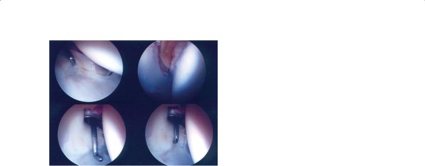
Figure 5.5 Arthroscopic images of a hip. The image at the top left shows a small acetabular labral tear
Ischial bursitis—The ischial bursa separates the gluteus maximus from the ischial tuberosity. Bursitis can arise from prolonged sitting or trauma to the bursa (hence the name “weaver’s bottom”). Use of a cushion and local corticosteroid injection may be useful.
Meralgia paraesthetica—This condition refers to local compression of the lateral cutaneous femoral nerve (L2-3 distribution) at the inguinal ligament. Patients experience numbness and burning pain in the anterior thigh. The syndrome is felt to arise from direct compression of the nerve; hence, obesity, pregnancy, tight-fitting belts and waistbands and hip extension (as can occur with high heels) are risk factors, as is diabetes mellitus. The syndrome generally improves with conservative measures such as weight loss, and changes in clothing and shoes.
Torn acetabular labrum—This produces pain in the groin on rotatory movements of the hip, and the hip may feel unstable or give
way. Labral tears can be associated with deformity of the femoral head or acetabulum. An MRI shows the abnormality, and the torn labrum can be removed arthroscopically (Figure 5.5).
Management of hip pain
The most important step in management of the painful hip is to establish the underlying aetiology and to treat it as specifically as possible. Thus infection of the hip should be diagnosed expeditiously and treated with surgical drainage and prolonged parenteral antibiotics. Fractures should be diagnosed and stabilized. Inflammatory arthritis can be treated with systemic therapy.
Here we present a few general principles that apply to the management of hip pain due to any number of aetiologies. First, a cane can be extremely helpful in unloading the painful hip and relieving pain. Patients must be shown the proper use of the cane in the contralateral hand.
Second, as with most other joints, the hip can become stiff with disuse and develop flexion contractures. This can be avoided with gentle range-of-motion exercises. If patients are losing motion, referral to a physiotherapist can be helpful.
Finally, it is important to recognize that one musculoskeletal problem can lead to another. Patients with spinal stenosis frequently develop trochanteric bursitis, for example. So while it is tempting to make a single, fully encompassing diagnosis in patients with musculoskeletal pain, the reality is that more than one condition could be present. While a patient’s underlying problem may lie in the back, injection of a secondarily involved trochanteric bursa may provide dramatic benefit.
Further reading
McRae R. Clinical Orthopaedic Examination. Churchill Livingstone, Edinburgh, 1997.
Miller MD, ed. Review of Orthopaedics. WB Saunders, Philadelphia, 2000. Solomon L, Nayagam D, Warwick D. Apley’s System of Orthopaedics and
Fractures. 8th edn. Arnold, London, 2001.