Материал: ABC-of-Rheumatology-ABC-Series-

CHAPTER 6
Pain in the Knee
Adrian Dunbar1 and Mark Wilkinson2
1Skipton, North Yorkshire, UK
2University of Sheffield, Sheffield, UK
OVERVIEW
•Knee pain is a frequent presenting complaint in primary care.
•Knee pain may arise from overuse injuries, trauma, degenerative change and inflammatory conditions.
•Osteoarthritis and rheumatoid arthritis affect the knee commonly.
•In most cases knee pain responds to simple measures such as lifestyle modification, simple analgesia and physiotherapy.
•Pain poorly controlled by simple measures, mechanical symptoms such as instability and locking, and progressive disability are indications for referral to secondary care.
•Where infection or tumour are suspected as a cause of knee pain, urgent referral to secondary care is required.
The knee is the largest joint in the body. It is a complex hinge that is made up of two separate articulations: the tibio-femoral joint and the patello-femoral joint. Knee motion occurs in a complex manner involving three planes, although the vast majority of its motion occurs in the sagittal plane (from full extension through to 140 ° of flexion).
Pain in the knee joint is one of the most common musculoskeletal complaints that presents to primary care physicians, and may arise from a broad range of pathologies. In the younger patient, pain most commonly arises from sporting or overuse injuries, which may affect the intra-articular or extra-articular structures of the knee. The knee is also a common site for inflammatory and infective pathologies. In the older patient, the most common cause is degenerative disease. Knee pain arising from osteoarthritis is a major cause of disability in the older patient, the prevalence and health-care costs of which continue to rise as the population ages.
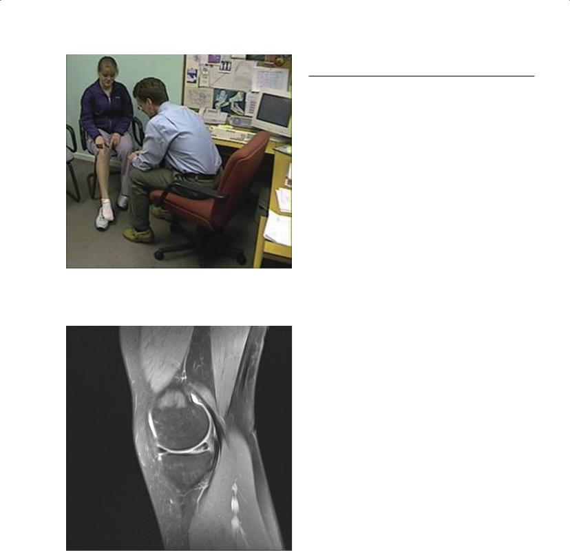
The evaluation of knee pain centres on a thorough history and physical examination supplemented, where necessary, with appropriate imaging and laboratory tests (Figure 6.1).
ABC of Rheumatology, 4th edn. Edited by Ade Adebajo. ©2010 Blackwell Publishing Ltd. 9781405170680.
Traumatic causes of knee pain
Injuries are a common cause of knee pain. Most knee injuries in sport occur as a result of indirect trauma, such as a twisting moment to the knee. The structures most commonly injured by this mechanism are the menisci, the collateral ligaments and the cruciate ligaments. These structures may be damaged in isolation, or may occur in combination (for example the anterior cruciate ligament, medial collateral ligament and medial meniscus may be injured in O’Donoghue’s triad). Direct trauma to the knee (such as during contact sport, an industrial accident or a motor-vehicle collision) most commonly causes bone contusions, fracture or dislocation that may affect the patello-femoral or tibio-femoral joint. Dislocation of the tibio-femoral joint indicates high-energy trauma, and is commonly associated with neurovascular damage.
Meniscus injury
Meniscus injury in young people can present as an acute injury or as a chronic condition with an insidious onset. The majority of meniscus tears in young people occur after mildto moderateenergy twisting injuries and are typically isolated injuries or associated with a collateral ligament strain. The medial meniscus is damaged three times more commonly than the lateral meniscus (Figure 6.2). Higher-energy twisting injuries are commonly associated with an anterior cruciate ligament injury, an acute haemarthrosis and inability to bear weight. Patients with meniscus tears have focal tenderness over the joint line and may experience mechanical catching and locking symptoms in the knee in addition to joint effusion and pain. Magnetic resonance imaging (MRI) can aid in establishing the diagnosis in cases where the history and physical examination are equivocal. Acute tears that occur in the well-vascularized peripheral portion of the meniscus are amenable to arthroscopic repair, which preserves meniscus function. Where an anterior cruciate ligament injury is also present this is reconstructed concurrently. Chronic meniscal tears are typically avascular with degenerative characteristics and will not heal if repaired. Arthroscopic resection is confined to the torn and degenerate portions of meniscus, as early-onset osteoarthritis of the knee commonly follows complete meniscal resection.
Articular cartilage injury
Articular cartilage injury is often the result of a traumatic episode
32
Pain in the Knee |
33 |
|
|
|
|
Figure 6.1 A detailed history and examination are required to make an accurate clinical diagnosis in the patient presenting with knee pain
Figure 6.2 MRI of meniscus injury (sagittal view). The anterior part of the medial meniscus can be seen as a black triangle on the left side of the joint line; the black triangle of the posterior part of the meniscus has a white line running through it, representing an oblique tear
that involves an impact injury to the cartilage surface. Articular cartilage injuries can result in focal pain, joint effusion and mechanical catching symptoms. Treatment comprises graduated physiotherapy for undisplaced injuries and arthroscopic repair or
Table 6.1 Post-traumatic knee swelling and the most common associated diagnoses
Immediate |
Delayed effusion |
Minimal effusion |
haemarthrosis |
|
|
|
|
|
Anterior cruciate |
Meniscus tear |
Collateral ligament tear |
ligament tear |
|
|
Osteochondral fracture |
Posterior cruciate |
|
|
ligament tear |
|
Patellar dislocation |
|
|
|
|
|
removal for displaced osteochondral fragments. Occult episodes of trauma to the knee may result in separation of cartilage from the subchondral bone, termed osteochondritis dissecans. Patients complain of poorly localized pain. The diagnosis is made from plain radiographs or MRI scans, and treatment commonly involves arthroscopic resection of loose cartilage.
A detailed history of the mechanism of injury and physical examination provide valuable information to differentiate between the various traumatic causes of knee pain. Knee pain from injury has a sudden onset at the time of the injury episode and is often accompanied by local soft-tissue swelling and an effusion. Certain fractures and dislocations may exhibit gross deformity; however, the majority of knee and patellar dislocations spontaneously reduce before presentation. A haemarthrosis develops quickly (over a period of minutes to a few hours) and indicates significant intraarticular injury, such as an anterior cruciate ligament tear, intraarticular fracture or osteochondral injury, or patellar dislocation. Effusions, which develop over several hours, tend to be associated with meniscal injuries (Table 6.1).
Radiographs should be obtained when evaluating any knee injury to exclude a fracture, dislocation or other significant abnormality. After obtaining radiographs, additional diagnostic tests may be indicated, including a computed tomography scan in the case of intra-articular fractures, or MRI when a soft-tissue or osteochondral injury is suspected. In the absence of neurovascular compromise or gross deformity, initial treatment of traumatic knee pain should consist of restricted weight bearing, ice and elevation. Severe injuries require immediate referral for orthopaedic surgical evaluation.
Knee pain in younger people and athletes
Knee pain in younger people and athletes can be caused by overuse syndromes, meniscus injury or articular cartilage abnormality. Common overuse syndromes include patellar tendonopathy, anterior knee pain syndrome, pes anserine bursitis and iliotibial band friction syndrome (Table 6.2).
Patellar tendonopathy
Patellar tendonopathy is caused by repetitive activity, particularly “explosive” athletics such as jumping. Patients complain of pain and soft-tissue swelling about the patellar tendon, usually at its

34 ABC of Rheumatology
Table 6.2 Symptoms associated with overuse injuries
Symptom |
Likely diagnosis |
|
|
Pain adjacent to patella |
Anterior knee pain syndrome |
Pain ascending/descending stairs |
|
Pain when sitting for prolonged periods |
|
(“movie theatre sign”) |
|
Pain in patellar tendon |
Patellar tendonopathy |
Pain with jumping |
|
Lateral knee pain with repetitive activity |
Iliotibial band friction syndrome |
Medial knee pain distal to joint line |
Pes anserine bursitis |
|
|
proximal attachment to the patella. Treatment consists of ice, painrelieving medication, activity modification and strengthening exercises focusing on eccentric loading of the tendon.
Anterior knee pain syndrome
Anterior knee pain syndrome occurs in patients who engage in repetitive athletic activity, in those with abnormalities in extensor mechanism alignment and in those who are overweight. Patients with anterior knee pain syndrome complain of pain in the front of the knee, which is accentuated by ascending and descending stairs, squatting, kneeling and by sitting for long periods of time. The pain may be located directly behind the patella or in the medial or lateral retinaculum. Treatment should include activity modification, weight control if necessary, physiotherapy to strengthen the quadriceps muscles (particularly vastus medialis) and core musculature, and appropriate pain-relieving medication.
Pes anserine bursitis
Pes anserine bursitis is an inflammation of the bursa overlying the insertion site of the semitendinosus, gracilis and sartorius tendons in the anteromedial aspect of the proximal tibia. Patients complain of medial knee pain distal to the medial joint line. Treatment can include activity modification, strengthening exercises and antiinflammatory medication. Chronic symptoms may respond to local corticosteroid injection.
Iliotibial band friction syndrome
Iliotibial band friction syndrome is an inflammation of the iliotibial band, the distal portion of the tensor fascia lata muscle that inserts into the anterolateral aspect of the proximal tibia. Patients are usually runners or cyclists who complain of activity-related lateral knee pain. This condition responds well to activity modification, stretching and strengthening exercises, ice and anti-inflam- matory medications.
Knee pain in older people
Twenty-five percent of people over the age of 50 report chronic knee pain, and degenerative arthritis of the knee is common in this
Box 6.1 Diagnosis of osteoarthritis
•Osteoarthritis is diagnosed clinically by the presence of:
Chronic knee pain
Morning stiffness lasting less than 30 minutes
Joint crepitus
Range of movement restricted by pain
Presence of osteophytes
Box 6.2 Non-pharmacological treatments
•Non-pharmacological treatments with an evidence base include:
Weight loss
Aerobic exercise
Specific knee-strengthening exercise
Patellar taping
Acupuncture
Knee bracing
age group (Box 6.1). However, clinical symptoms and radiological severity of arthritis are poorly correlated. Many older people with knee pain have minor radiological evidence of arthritic change. Conversely, many people with advanced radiological changes are pain-free. Arthritis of the knee is often associated with periarticular soft-tissue problems, and indeed these can often be a major source of knee pain. Pes anserine bursitis is a common example. Plain radiographic imaging is not always helpful in the assessment of patients with knee pain, and the diagnosis of osteoarthritis is often a clinical one. An MRI scan may assist in the diagnosis of an occult degenerate meniscal tear.
The management of osteoarthritis is, for most people, the management of their knee pain and lifestyle modification (Box 6.2). The high prevalence of knee pain in the community means that such treatments should be simple, safe, cost-effective and, ideally, self-administered. Initial treatments consist of simple analgesia, such as paracetamol, that is safe and effective. The place of oral glucosamine and similar nutraceuticals is still debated in the presence of conflicting reports from different studies, and none have yet been convincingly shown to alter the course of osteoarthritis.
Oral non-steroidal anti-inflammatory drugs (NSAIDs) are prescribed commonly; however, there is little evidence of benefit over simple analgesia, and they are associated with significant risk of serious adverse effects in the older patient. Their use should be considered after failure of simple measures such as weight loss, exercise regimes and use of simple analgesics.
Local treatments, such as topical non-steroidal anti-inflamma- tory gels are effective in the short term, particularly in the setting of acute symptomatic flares. Injected treatments include corticosteroids and hyaluronans. Intra-articular steroids can be very effec-
Pain in the Knee |
35 |
|
|
|
|
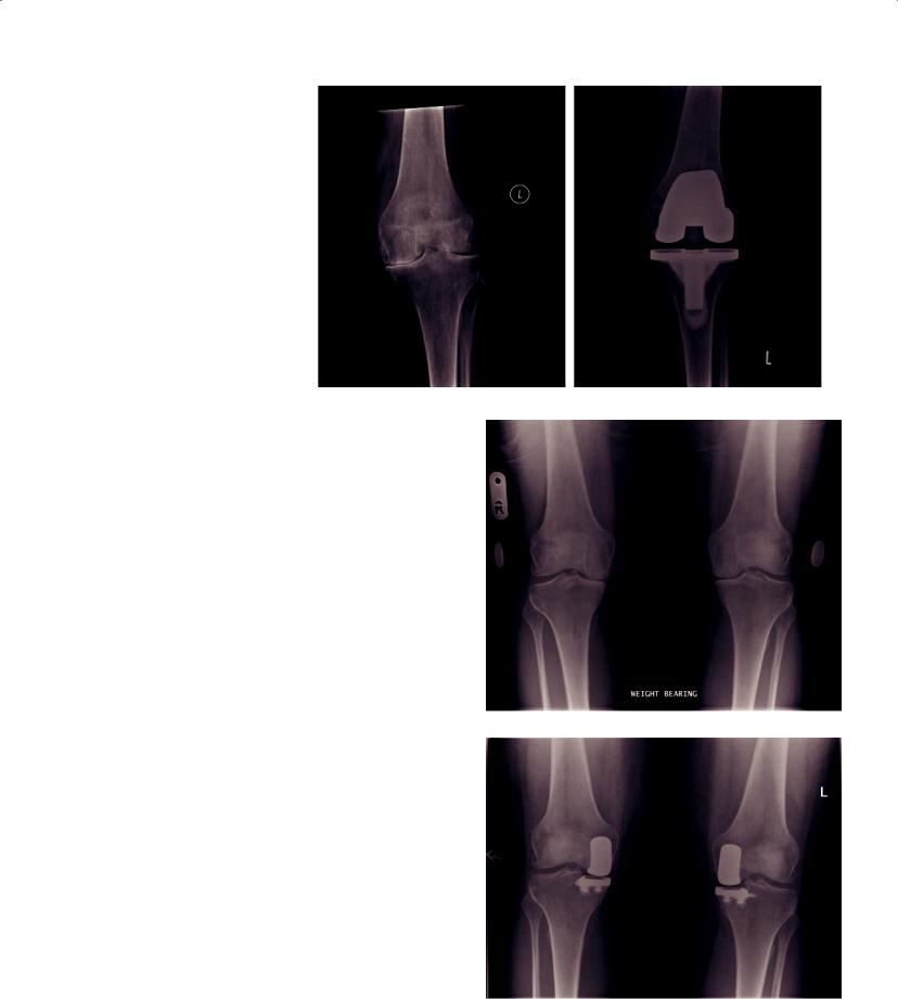
Figure 6.3 Radiograph showing the |
|
|
typical features of knee osteoarthritis, |
|
|
joint-space narrowing, subchondral |
|
|
sclerosis and osteophyte formation |
|
|
(a), and following treatment with |
(a) |
(b) |
total knee replacement (b) |
tive in relieving knee pain for several weeks or months. Hyaluronans have a longer-lasting effect, but are very much more expensive and require a series of injections over time. Both have good safety profiles, although certain hyaluronans can cause pseudoseptic joint inflammation and effusion.
Arthroscopic surgical treatment for arthritis of the knee is reserved for the treatment of mechanical symptoms such as joint catching, locking or instability due to a loose body or meniscal tear. In the absence of mechanical symptoms, arthroscopic interventions are no more effective than placebo.
In up to 40% of patients, disease does not progress significantly after initial presentation, or does so very slowly. In these patients use of simple, safe, cost-effective treatments is essential for effective and economic management. Joint replacement surgery is indicated in those patients whose disease progresses such that their symptoms become poorly controlled despite the treatment measures outlined above. In most patients this entails total knee replacement (Figure 6.3). In a small proportion of patients the arthritis is limited to one compartment of knee, in which case a unicompartmental joint replacement is an effective alternative to total knee replacement, and is associated with good functional outcomes in suitable patients (Figure 6.4). The results of joint-replacement surgery are excellent in over 90% of patients in terms of improvement in health-related quality of life.
Knee pain in systemic disease
Pain and swelling in the knee may be a feature of systemic illness. Patients should be asked about pain in other joints, previously painful, swollen joints and a family history of joint disease. Systemic symptoms such as malaise, pyrexia, anorexia and weight loss may provide clues to the origin of the knee pain. Symptoms affecting other organs, such as the skin, bowel, eyes or genito-urinary tract, may also be of diagnostic relevance.
(a)
(b)
Figure 6.4 Radiographs showing bilateral isolated osteoarthritis of the medial compartment of the knee (a), and following treatment with bilateral unicompartmental knee replacement (b)
36 ABC of Rheumatology
The knee is the most commonly affected large joint in rheumatoid arthritis. The knees are usually affected bilaterally, and symptom onset usually occurs early in the course of the disease. The knee is also commonly affected in the other chronic inflammatory arthritides, including psoriatic arthritis and ankylosing spondylitis. The treatment of the knee pain in these conditions is considered along with the management of the systemic disease and includes lifestyle modification, physiotherapy, disease-modifying agents, NSAIDs, novel biological agents and total joint-replace- ment surgery.
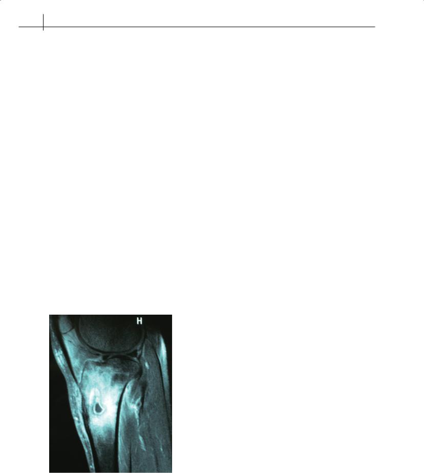
The knee is the most commonly infected joint. Joint infection presents with a red, swollen, hot knee, difficulty in weight bearing and a limitation in the range of passive motion. Occasionally, the infection may originate in the metaphyseal region of the tibia or femur, rather than the knee joint itself (Figure 6.5). A suspected infection of the knee requires immediate referral to secondary care for assessment and treatment. The most common infecting organism is Staphylococcus aureus. Less common infections include
Streptococcus, Gonococcus, Brucella and, rarely, tuberculosis. Infective arthritis should always be considered in the immunocompromised and other patients with increased infective risk, e.g. intravenous drug users.
Aspiration of the joint for microbiological culture is the most important investigation for the accurate diagnosis of infection. This must be carried out at initial assessment, and before the administration of antibiotics. Aspiration of the knee made after antibiotic administration often results in a false-negative microbiological culture result and a missed diagnosis. Other useful diagnostic tests include concurrent aspirate microscopy for crystals, and
serological measurement of white cell count, erythrocyte sedimentation rate and C-reactive protein. The treatment of the infected knee includes initiation of systemic antibiotics immediately after knee aspiration, typically using an agent with broad Gram-positive antimicrobial activity, and serial joint aspiration or arthroscopicassisted washout. The choice of antibiotic is adjusted as indicated by the aspirate microbiological culture sensitivities, and may be continued for up to 6 weeks orally, although specialist microbiological advice should be taken where infection is confirmed from aspirate culture.
The differential diagnosis of the hot, swollen, painful knee includes systemic inflammatory conditions such as calcium pyrophosphate arthropathy, gout, Reiter’s disease and pre-patellar bursitis. Aspiration of joint fluid for crystal microscopy and culture is important, as are appropriate serological investigations, both in confirming the correct diagnosis and in excluding joint infection. Rarely, infections of the genito-urinary tract and viral infections may present with bilateral swollen, tender knees with a large effusion of sympathetic origin. Radiographs are frequently of limited diagnostic utility in such cases.
Other causes of knee pain
Hip pain may occasionally refer to the anterior distal thigh or the knee. A complete examination of the patient with knee pain includes an examination of the hip to exclude this cause of knee pain. Knee pain may also present as part of a chronic widespread pain syndrome. An adequate general musculoskeletal assessment is essential if appropriate treatment of the knee pain is to be effected. In the presence of polyarthralgia, or symptoms suggestive of a fibromyalgia syndrome, the knee pain is unlikely to be adequately managed by focusing on the knee alone. Attention should be paid to management of the global pain problem.
Figure 6.5 MRI scan of the knee (sagittal view) in a patient presenting with an acute, red knee. In this case the diagnosis was acute Staphylococcal osteomyelitis of the proximal tibial metaphysis
“Red flags”
Although primary bone tumours are rare, the knee is one of the most commonly affected sites for benign tumours, including osteoid osteoma, enchondroma and chondroblastoma, and malignant tumours, including osteosarcoma and chondrosarcoma. Ewing’s sarcomas also commonly affect the knee. In children and young adults who are very active, knee pain may be related to recent activity. Unexplained pain, pain that is worse at night, unexplained swelling and systemic symptoms are all “red flag” features that may indicate a bone tumour. Patients in whom a bone tumour is suspected should be referred early to a centre specializing in their management.
Further reading
Brukner P, Khan K. Lateral, medial and posterior knee pain. In: Brukner P & Khan K, eds. Clinical Sports Medicine, 3rd edn. McGraw Hill, London, 2007. Available online at http://www.clinicalsportsmedicine.com/ chapters/index.htm
Brukner P, Khan K, Cooper R, Morris H, Arendt, L. Acute knee injuries. In: Brukner P & Khan K, eds. Clinical Sports Medicine, 3rd edn. McGraw Hill,