Материал: ABC-of-Rheumatology-ABC-Series-
22 ABC of Rheumatology
Nucleus pulposus
 Annulus fibrosus
Pedicle
Cauda equina
Lamina |
Facet joint |
|
Ligamentum flavum
Spinous process
(a)
Zygapophyseal |
Intervertebral |
|
disc |
||
joint |
||
|
||
(Facet joint) |
|
Intervertebral foramina
(b)
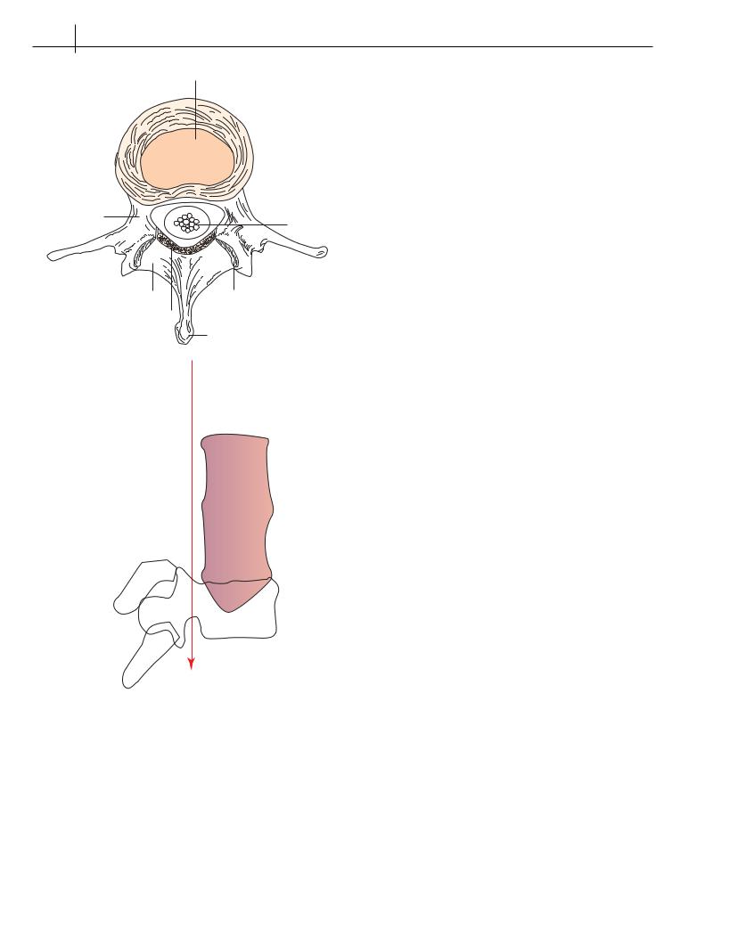
Figure 4.1 Basic anatomy of the lumbar spine; (a) cross-sectional view through a normal lumbar vertebra; (b) lateral view of the lumbar spine
herniations, 95% involve the L4-5 or L5-S1 disc. Generally, the more caudal nerve root is impinged; that is, the L5 nerve root with L4-5 herniation and S1 nerve root with L5-S1 herniation. In most patients the sciatic pain resolves over a period of weeks.
Rarely, a large midline disc herniation compresses the cauda equina. This is a surgical emergency. The full cauda equina syndrome usually presents with bilateral sciatica and motor deficits. It
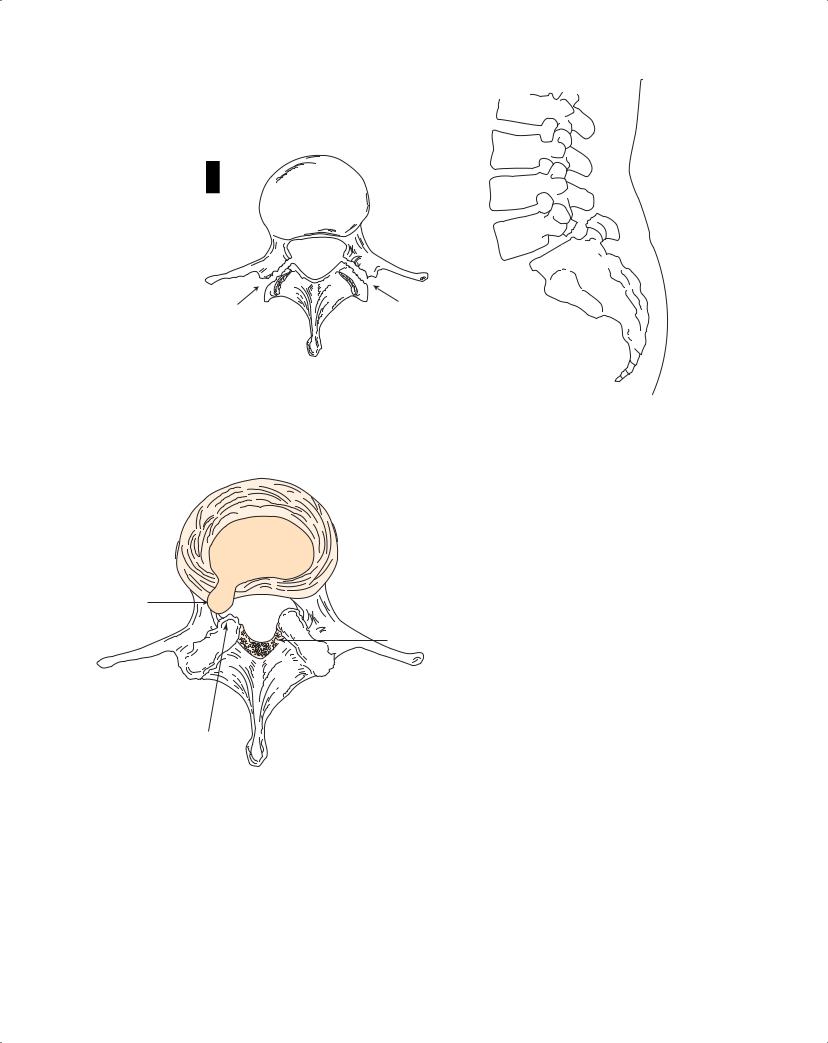
Figure 4.2 Posterolateral disc herniation resulting in nerve root impingement
Box 4.2 Common symptoms of cauda equina syndrome
The patient will develop some or all of the following:
•Altered saddle or/and urinary sensation
•Rectal/perineal pain
•Change/reduced awareness of bladder filling
•Need to strain to maintain urine flow
•Difficulty in walking or the legs just “do not feel right” (very early symptoms)
•Urinary retention with overflow incontinence and faecal incontinence (late manifestations of the full syndrome)
needs to be recognized and treated before urinary retention and/ or incontinence occur for a completely successful outcome. Common symptoms are presented in Box 4.2.
Spondylolisthesis
Spondylolisthesis is the anterior displacement of a vertebra on the one beneath it. It is usually secondary to degenerative changes in the disc and facet joints (degenerative spondylolisthesis) but may result from a developmental defect in the pars interarticularis of the vertebral arch (spondylolysis), which produces isthmic spondylolisthesis (Figure 4.3). Patients with minor degrees of spondylolisthesis are usually asymptomatic, although some may have mechanical LBP. Greater degrees of spondylolisthesis occasionally cause sciatica or spinal stenosis.
Spinal stenosis
Spinal stenosis (ST) is defined as a narrowing of the spinal canal and its lateral recesses and neural foramina, which may result in a
Low Back Pain |
23 |
|
|
|
|
L5
S1
(a) |
(b) |
Figure 4.3 (a) Spondylolysis with bilateral defects in the pars interarticularis (arrows); (b) spondylolysis at L5 resulting in spondylolisthesis at L5-S1
A
C
B
Figure 4.4 Spinal stenosis secondary to a combination of disc herniation (A), facet-joint hypertrophy (B) and hypertrophy of the ligamentum flavum
(C)
compression of lumbosacral nerve roots (20% of adults over age 60 have imaging evidence of ST but are asymptomatic).Degenerative changes (leading to disc herniation, facet joint osteophytes and ligamentum flavum hypertrophy) are the causes of ST in most patients (Figure 4.4).
The hallmark of ST is pseudoclaudication (neurogenic claudication). Symptoms are often bilateral with pain, weakness and some-
times paraesthesiae in the buttocks, thighs and legs. Symptoms are induced by standing or walking and relieved by sitting or flexing forward. Forward flexion increases the canal diameter and may lead to the adoption of a simian stance. Unsteadiness of gait is common. Physical examination is usually unremarkable, and severe neurologic deficits are rarely seen. The diagnosis is best confirmed by magnetic resonance imaging (MRI).
Idiopathic low back pain
A definitive pathoanatomical diagnosis with precise identification of the pain generator cannot be made in 80% of patients. Nonspecific terms such as lumbago, strain and sprain (which have never been anatomically or histologically characterized) have come into use for this mostly self-limited syndrome of LBP.
Assessment
A major focus of the evaluation is to identify the few patients with an underlying systemic disease (infection, neoplasm or spondyloarthropathy) or significant neurologic involvement that may require urgent and/or specific intervention. It is essential to take a full history and perform a comprehensive physical examination.
History
The patient’s back pain should be characterized. Severe mechanical LBP with an acute onset in a slender postmenopausal woman is suspicious for a vertebral compression fracture secondary to osteoporosis. Non-mechanical LBP, especially when accompanied by nocturnal pain, suggests the possibility of underlying infection or neoplasm. Inflammatory LBP, as seen in the spondyloarthropathies, is accompanied by night-time waking with pain and stiffness
24 ABC of Rheumatology
and/or prolonged morning stiffness that improves with exercise but not with rest. The radicular pain of sciatica suggests nerve root impingement. It should be differentiated from non-neurogenic sclerotomal pain. Pseudoclaudication is seen with spinal stenosis.
Physical examination
This rarely leads to a specific diagnosis. Inspection may reveal a structural or functional scoliosis. Structural scoliosis is secondary to structural changes of the vertebral column. Functional scoliosis
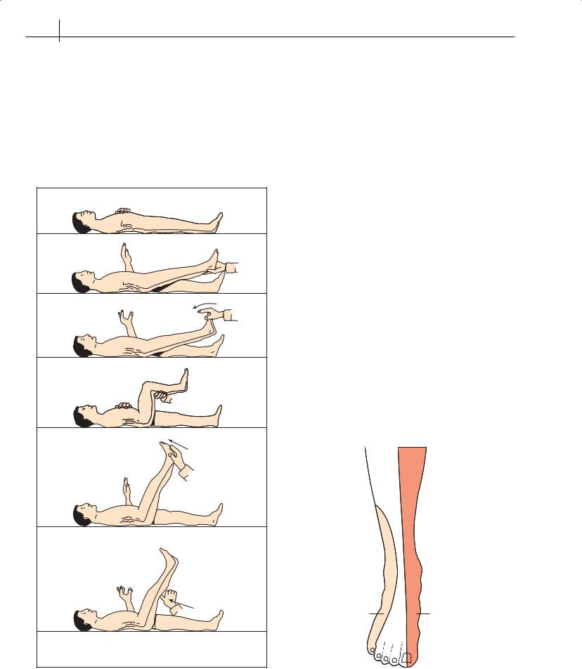
(a) when supine, the nerve roots (L4, L5 and S1) are slack.
(b) Straight leg raising is limited by the tension of the root over the prolapsed disc.
(c) Tension is increased by dorsiflexion of the foot.
(d) Root tension is relieved by flexion at the knee and ankle.
(e) With the knee extended, the root tightens over the prolapsed disc causing pain which radiates to the back.
is usually the result of paravertebral muscle spasm or leg-length discrepancy. Functional scoliosis disappears with spinal flexion, whereas structural scoliosis persists.
Paravertebral muscle spasm often leads to loss of the normal lumbar lordosis. Point tenderness on percussion over the spine has sensitivity but not specificity for vertebral osteomyelitis. A palpable step-off between adjacent spinous processes indicates spondylolisthesis.
Limited spinal motion is not associated with any specific diagnosis, because LBP due to any cause may limit motion. Range-of- motion measurements can help in monitoring treatment. Examine the hip for arthritis: this normally causes groin pain, and occasionally referred back pain.
A straight leg raise test (Figure 4.5) should be performed on all patients with back pain that radiates into the lower extremities. This test places tension on the sciatic nerve and stretches the sciatic nerve roots (L4, L5, S1, S2 and S3). Patients with existing nerve root irritation, e.g. impingement from a herniated disc, will experience radicular pain that extends below the knee. This test is very sensitive (95%) but not specific (40%) for clinically significant disc herniation at the L4-5 or L5-S1 level. The straight leg raise test is usually negative in patients with spinal stenosis.
For lower extremities, neurologic evaluation should include motor testing, determination of knee and ankle deep tendon reflexes, and dermatomal sensory loss tests (Figure 4.6). This can help identify the specific nerve root involved (Table 4.1), e.g. a significant left-sided L5-S1 posterolateral disc herniation often impinges upon the left S1 nerve root. Patients will have left-sided sciatica in the distribution of the S1 dermatome and may develop left plantar flexion weakness, diminished light touch and pinprick sensation over the lateral aspect of the foot, and a diminished or absent left ankle jerk.
S1 |
L5 |
L4 |
(f) Pressure over the centre of the popliteal fossa pulls on the posterior tibial nerve which is ‘bow stringing’ across the fossa causing local pain and radiation to the back.
Figure 4.5 Examination for pain in the back: supine position |
Figure 4.6 Lower-extremity dermatomes |
Low Back Pain |
25 |
|
|
|
|
Table 4.1 Neurological features of lumbrosacral radiculopathy
Disc |
Nerve |
Motor |
Sensory |
Reflex |
herniation |
root |
|
(light touch) |
|
|
|
|
|
|
L3-4 |
L4 |
Dorsiflexion |
Medial foot |
Knee |
|
|
of foot |
|
|
L4-5 |
L5 |
Dorsiflexion |
Dorsal foot |
None |
|
|
of great toe |
|
|
L5-S1 |
S1 |
Plantar flexion |
Lateral foot |
Ankle |
|
|
of foot |
|
|
|
|
|
|
|
Box 4.3 “Red flags” that indicate need for early diagnostic testing
Spinal fracture
•Significant trauma
•Prolonged glucocorticoid use
•Age >50 years
Infection or cancer
•History of cancer
•Unexplained weight loss
•Immunosuppression
•Injection drug use
•Nocturnal pain
•Age >50 years
Cauda equina syndrome
•Urinary retention
•Overflow incontinence
•Faecal incontinence
•Bilateral or progressive motor deficit
•Saddle anaesthesia
Spondyloarthropathy
•Night-time waking with pain and stiffness
•Morning stiffness in the back
•Low back pain that improves with activity
•Age <40 years
Imaging studies
Diagnostic testing is rarely indicated unless symptoms persist beyond 4 weeks, as 90% of patients will have recovered within this time, thus avoiding unnecessary testing. “Red flags” indicate early investigations, e.g. underlying systemic disease or patients with a significant neurologic deficit (Box 4.3).
A major problem with all imaging studies is that many of the anatomical abnormalities (often the result of age-related degenerative changes) are common in asymptomatic people. Abnormalities such as single disc degeneration, facet-joint degeneration, Schmorl’s nodes, spondylolysis, mild spondylolisthesis, transitional vertebrae (lumbarization of S1 or sacralization of L5), spina bifida occulta
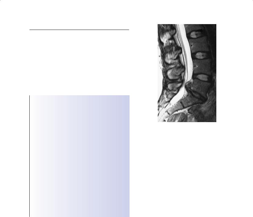
Figure 4.7 MRI showing a posterolateral disc prolapse
and mild scoliosis are equally prevalent in people with and without LBP. Plain radiographs are usually unhelpful in determining the cause of LBP and should be limited to patients with findings suggestive of systemic disease (infection, neoplasm, spondyloarthropathy) or trauma, or those with continued LBP after 4–6 weeks of conservative care.
Computed tomography and MRI (Figure 4.7) should be reserved for patients in whom underlying infection or cancer is suspected, or for patients with significant or progressive neurologic deficits. MRI is the preferred modality for the detection of spinal infection, neoplasm, herniated discs and spinal stenosis. Bone scanning is used primarily to detect bony metastases, occult fractures and infection.
Treatment
Most patients, regardless of the cause, respond to a general programme that includes analgesia, education, back exercises, aerobic conditioning and weight control. Specific treatment is available only for the small number of patients with major neurologic compression or underlying systemic disease.
For treatment purposes, patients are considered to have acute LBP (duration <3 months), chronic LBP (duration >3 months) or a nerve root compression syndrome.
Acute LBP
Patients are advised to stay active, and bed rest is discouraged. Acetaminophen/paracetamol and non-steroidal anti-inflammatory drugs (NSAIDs) offer symptomatic relief; some people will need short-term narcotic analgesics, and muscle relaxants used for a few days will help others.
26 ABC of Rheumatology
Once the acute episode of pain has subsided, a programme of regular back exercises (including stretching), aerobic conditioning and loss of excess weight is used to prevent recurrences. Back exercises help to stabilize the spine. Flexion exercises strengthen the abdominal muscles and extension exercises the paraspinal muscles. Educational booklets that include back exercises and safe lifting techniques are helpful.
Many patients ask about chiropratic and osteopathy treatments. There is no evidence that spinal manipulative therapy is superior to standard treatment for back pain.
Chiropractic focuses on the diagnosis, treatment and prevention of mechanical disorders of the musculoskeletal system, and on the effects of these disorders on the nervous system and on general health. It was founded in the USA by DD Palmer in 1895 who based it on his belief that disorders are caused by misaligned vertebrae which cause nerve compression (subluxations) and dysfunction. The primary chiropractic technique being adjustment of the spine. Chiropractors may specialize in low back pain problems, or they may combine chiropractic with manipulation of the extremities, physiotherapy, nutrition or exercise to improve the strength of the spine.
The General Chiropractic Council (www.gcc-uk.org) has regulated the chiropractic profession since 1994.
Osteopathy is a manual therapy that is primarily focused on the treatment of musculoskeletal conditions. It was founded by Andrew Still in the USA in 1886, who believed that disease was caused when the flow of nerve impulses was disrupted when a person’s bones were out of place. He concluded that manipulating bones back into place would restore the interrupted flow of nerve impulses and cure disease.
Underpinning osteopathy is the idea that the body has self-regulatory mechanisms and therefore that it has the capacity to heal itself, that the structure and function of the body are closely inter-related, and that the somatic aspects of disease aren’t just manifestations of disease but also contribute to maintenance of the disease state. Osteopaths will commonly treat back pain by manipulation, but may also use soft, tissue massage or advise exercise.
Osteopathy was established in the UK in 1917, and has been subject to statutory regulation since 1993. It is regulated by the General Osteopathic Council (www. osteopathy.org.uk).
There is limited evidence supporting the use of epidural glucocorticoid injections for short-term relief of radicular pain. Nerveroot blocks and injection of anaesthetic agents or glucocorticoid into trigger points, ligaments, sacroiliac joints and facet joints are of unproven efficacy.
Ultrasound, shortwave diathermy, transcutaneous electrical nerve stimulation and other treatments such as lumbar braces, traction, acupuncture and biofeedback are ineffective.
Chronic LBP
Treatment of chronic LBP is focused on relief of pain and restoration of function. Complete relief of pain is an unrealistic goal for most. Acetaminophen/paracetamol and NSAIDs may provide some degree of analgesia. Long-term use of narcotic analgesics should be avoided. Low-dose tricyclic antidepressants may help some patients.
Back exercises, aerobic conditioning, loss of excess weight and patient education are effective in managing chronic LBP. A multidisciplinary approach focusing on functional restoration through an intensive rehabilitation programme based on cognitive behavioural therapy is often helpful.
The results of back surgery are disappointing when the goal is relief of back pain (such as by spinal fusion or artificial discs) rather than relief of radicular symptoms from neurologic compression.
Nerve root compression syndromes
Disc herniation—Patients with radicular pain secondary to nerveroot compression should be treated conservatively, as described for acute LBP, for the first 6 weeks unless there is severe or progressive neurologic deficit: approximately 90% will improve. Elective surgery may be considered in a few patients among those who have a significant persistent neurologic deficit or severe sciatica after 6 weeks of conservative care. Laminotomy with limited discectomy is generally the procedure of choice.
Spinal stenosis—The symptoms remain stable for years in most patients. Analgesics, NSAIDs, loss of excess weight, exercises (including those that reduce lumbar lordosis) and epidural glucocorticoids may provide symptomatic relief. Surgical treatment, aimed at decompression of the neural elements, is offered to patients with either disabling pseudoclaudication or significant neurologic deficit.
Further reading
Carragee EJ. Persistent low back pain. New England Journal of Medicine 2005; 352: 1891–1898.
Critchley D, Hurley M. Management of Back Pain in Primary Care. Reports on the Rheumatic Diseases Series 5, no. 13. Arthritis Research Campaign, York, UK, 2007. Available online at http://www.arc.org.uk/arthinfo/ medpubs/6533/6533.asp
Deyo, RA, Weinstein JN. Low back pain. New England Journal of Medicine 2001; 344: 363–370.
HMSO. The Back Book. The Stationery Office, London, 2000.
Imboden J, Hellmann D, Stone J. Current Diagnosis and Treatment in Rheumatology, 2nd edn. McGraw-Hill, New York, 2007.
Jarvik JG, Deyo RA. Diagnostic evaluation of low back pain with emphasis on imaging. Annals of Internal Medicine 2002; 137: 586–597.
Shapiro S. Medical realities of Cauda equine syndrome secondary to lumbar disc herniation. Spine 2000; 25: 348–351.