Материал: ABC-of-Rheumatology-ABC-Series-
Pain in the Neck, Shoulder and Arm |
17 |
|
|
|
|
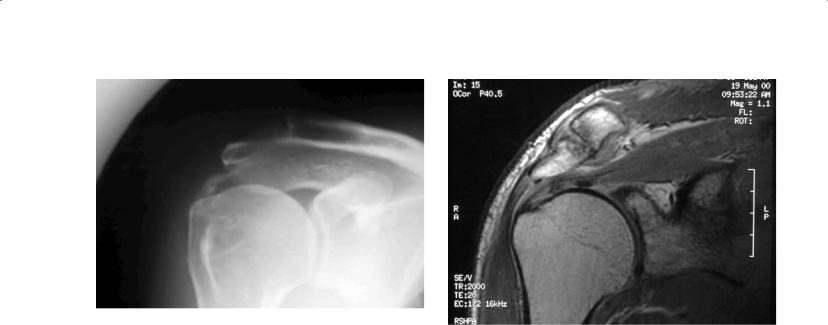
Figure 3.6 Plain X-ray showing calcific tendinitis. Adapted from Speed |
|
et al., 2000 |
Figure 3.7 MRI scan of shoulder, showing acromioclavicular osteophytes |
|
and underlying rotator cuff tendinitis |
Treatment—The aims of treatment of rotator cuff disease are to control pain and restore movement and function of the shoulder. Paracetamol is suitable as first-line therapy and may be supplemented by mild opioids such as codeine phosphate if needed. NSAIDs may provide short-term pain relief if there are no contraindications to their use. Initially patients may need to modify their activities and address occupational factors.
Subacromial injection of depot corticosteroid and local anaesthetic may provide rapid relief of pain, but its effect may be small and not maintained beyond a few weeks. If initial response is good, injections may be repeated up to two or three times at six-weekly intervals. Although injections performed under fluoroscopy or ultrasound might increase the accuracy of needle placement, it is not clear whether or not this results in significantly better outcomes.
Physiotherapy comprising a combination of mobilization techniques and directed exercises designed to strengthen and stabilize the cuff and scapular muscles can be used alone or combined with other measures. Global strengthening and proprioception training may reduce instability and minimize impingement in those with glenohumeral joint hypermobility.
Benefits of heat or ice packs, low-power laser, ultrasound and pulsed electromagnetic field therapy are unproven, as trials have yielded conflicting results. There is limited evidence for transitory pain relief following acupuncture, and suprascapular nerve block may also provide short-term pain relief. Trials have failed to establish the efficacy of extracorporeal shock wave therapy (ESWT) for rotator cuff disease.
Surgery may be required when symptoms fail to respond to conservative treatment. Operative treatment involves decompression of the subacromial space, with or without rotator cuff repair. MRI may be useful to plan surgery (Figure 3.7). Observational studies have reported good outcomes of surgery, although three randomized controlled trials found that surgery was not superior to treatment with supervised exercises.
Subacromial steroid injections, needling of the calcific deposits under fluoroscopic guidance and percutaneous needle aspiration and lavage by ultrasound guidance have each been advocated to relieve pain in calcific tendinitis, although no data are available from controlled trials. Ultrasound may provide short-term pain relief and, like ESWT, may improve the radiological appearance of calcific deposits. Surgical removal of calcific deposits may be of benefit if conservative treatments fail.
Adhesive capsulitis
Adhesive capsulitis (“frozen shoulder”, or painful stiff shoulder) affects 2–5% of the population, women slightly more often than men, and 10–36% of people with diabetes, in whom it is more severe. It occurs most commonly in the fifth and sixth decades of life and is rare before the age of 40 years. The cause is poorly understood. It is usually idiopathic, although it may occur in the context of prolonged shoulder immobility (e.g. following a stroke or cardiac, breast or shoulder surgery).
Three phases have been described: initial gradual development of diffuse and severe shoulder pain, typically worse at night with inability to lie on the affected side, lasting between 2 and 9 months; a stiff phase with less severe pain present at the end range of movement, characterized by global stiffness and severe loss of shoulder movement, lasting about 4 and 12 months; and finally a recovery phase characterized by a gradual return of movement over 5 and 24 months. Severe disability may result in absence from work and inability to perform leisure activities. Although generally thought to run a self-limiting course over 2 to 3 years, some studies have found that up to 40% of patients have persistent symptoms and restricted movement beyond 3 years.
Diagnosis—The diagnosis can be made clinically, as the restriction of both active and passive movement in all planes of movement, especially external rotation, distinguishes it from other causes of shoulder pain. Plain radiographs are not necessary in primary care

18 ABC of Rheumatology
unless glenohumeral arthritis is suspected. Likewise, MRI is seldom necessary to establish the diagnosis, even in specialist care.
Treatment—Treatment is needed to control severe pain, improve range of movement and promote function. Patients should be informed of the generally favourable prognosis.
Treatment with analgesia and NSAIDS is the same as for rotator cuff disease.
Intra-articular injection of corticosteroid combined with local anaesthetic using either an anterior or posterior approach may provide rapid pain relief, but the effect may not be sustained beyond 6–7 weeks. There are limited data to provide guidance about frequency, dose and type of corticosteroid for adhesive capsulitis.
Arthrographic distension of the glenohumeral joint (or hydrodilatation) is performed under radiological guidance, usually using a combination of local anaesthetic, corticosteroid and saline to a mean volume of 20–45 ml. It has recently been demonstrated to have a sustained beneficial effect on pain, function and range of movement and is the standard of care in some settings. It may be more effective in the intermediate (stiff) and recovery stages and may also be repeated if the effect wanes over time.
Physiotherapy in the early, painful phase of the condition may aggravate the pain. However, gentle mobilization and strengthening exercises can improve mobility and reduce the duration of disability in the later phases. There is also evidence that mobilization and strengthening exercises following either steroid injection or arthrographic distension provide additional benefits over these treatments alone.
A short course of oral glucocorticoids, prescribed by a specialist, may provide rapid pain relief, although the effect may diminish beyond 6 weeks. Although treatment may be more effective in the very early phase of the condition, benefit has been demonstrated in patients with an average duration of symptoms of 5 to 6 months.
Suprascapular nerve blocks may provide short-term pain relief. Manipulation under anaesthesia, possibly combined with intraarticular steroid injection and/or arthroscopic debridement of adhesions, may be helpful if conservative options have failed. Manipulation under anaesthesia can however, cause iatrogenic damage such as fractures, haemarthroses and tears of the labrum,
tendons or ligaments.
Other shoulder disorders
Acromioclavicular and sternoclavicular joint disorders—Osteoarthri- tis of the acromioclavicular joint is common and presents with well-localized pain and tenderness over the joint. It can be managed symptomatically with analgesics, and local corticosteroid injections may provide relief. Surgery can be effective in resistant cases. The acromioclavicular joint can be strained or dislocated as a result of traumatic or sports injuries. Examination may find a superior painful arc of abduction (Figure 3.5) and restriction of passive horizontal adduction (flexion) of the shoulder, with the elbow extended across the body. This can be managed with taping and analgesia, and in severe cases, surgery may be required.
The sternoclavicular joint can be the presenting site of an inflammatory arthritis, but it is frequently overlooked. Rarely the sternoclavicular and/or acromioclavicular joints can be involved in the
rare SAPHO syndrome (synovitis, acne, pustulosis, hyperostosis, osteomyelitis).
Glenohumeral joint arthritides—Isolated osteoarthritis of the shoulder is rare but may occur following fractures of the humeral head or neck or large rotator cuff tears, or as the end result of rheumatoid arthritis. It may be suspected, particularly in the older age group, if there is a limited range of painful movement sometimes accompanied by crepitus. Plain radiographs are useful in this instance. New onset of bilateral shoulder pain and stiffness should prompt consideration of polymyalgia rheumatica in those over 50 years of age and in rheumatoid arthritis. Milwaukee shoulder, which mainly affects elderly women, is a severe destructive apatiteassociated arthropathy that presents with shoulder pain, limited movements and large joint effusion. Aspiration reveals a large amount of blood-tinged synovial fluid, which contains calcium phosphate crystals.
Biceps tendinitis/rupture—The long head of the biceps tendon passes through the bicipital groove of the anterior proximal humerus and is often involved in rotator cuff disease but can present as an isolated problem. It presents with anterior shoulder pain, aggravated by lifting, carrying objects and overhead reaching. Sudden onset of worsening symptoms, which may occur after heavy lifting or be spontaneously accompanied by a swelling just above the antecubital fossa and sometimes bruising, suggests an acute rupture. In most instances proximal tendon rupture is cosmetic and does not require repair. However, distal biceps tendon rupture should be referred urgently for consideration of surgical repair.
Shoulder instability—General glenohumeral instability or looseness may be seen in young women with weak shoulder muscles, in young athletes (especially swimmers and throwers) and following large rotator cuff tendon tears. There may be diffuse shoulder pain, and instability may be multior unidirectional.
Glenoid labrum (cartilage) injuries—These can cause persistent shoulder pain and instability, and they usually occur after an episode of trauma or dislocation or with overuse. Diagnosis can be difficult, requiring magnetic resonance arthrography or arthroscopy. Management involves pain control and rehabilitation, which is followed by surgery if necessary.
Neurological causes—Shoulder pain may result from neurological causes, including nerve root entrapment at the neck, brachial plexus lesions or peripheral nerve lesions, including the axillary, long thoracic, suprascapular, radial or musculocutaneous nerves. Brachial neuritis can affect one or more components of the brachial plexus. Often idiopathic, some cases occur after a viral infection, immunizations or mechanical trauma. A sudden onset of diffuse pain in the shoulder, upper arm and occasionally forearm is accompanied by weakness, wasting, scapular winging and variable sensory loss of the affected neuromuscular structures. Electromyographic studies may be confirmatory. Tricyclic agents, carbamazepine, gabapentin or pregabalin may be helpful. Rehabilitation is started early to prevent stiffness and improve function.
Pain in the Neck, Shoulder and Arm |
19 |
|
|
|
|
Thoracic outlet syndrome—Compression of the neurovascular structures of the thoracic outlet, brachial plexus and subclavian artery may occur due to local masses, a high first or cervical rib or fibrous bands. Symptoms depend on the structures compressed, but they are usually exacerbated by heavy manual work. Neurogenic symptoms usually predominate, including aching in the arm, paraesthesia and weakness. Vascular symptoms are usually intermittent cyanosis; trophic skin changes can occur. A causative structure is rarely identified, and management is symptomatic.
Elbow and forearm pain
Lateral and medial epicondylitis
The 12-month period prevalence of elbow pain has been estimated to be 11.2%. Box 3.4 displays a list of differential diagnoses of elbow pain. Most complaints of elbow pain are due to lateral epicondylitis (“tennis elbow” or lateral elbow pain), which has an estimated annual incidence in general practice of 4–7 per 1000 patients. People aged between 40 and 50 years are most commonly affected. Lateral epicondylitis is thought to be an overload injury at the origin of the common extensors at the lateral epicondyle, and typically follows minor and often unrecognized trauma of the extensor muscles of the forearm. In spite of the title “tennis elbow”, tennis is a direct cause in only 5% of cases. Risk factors include repetitive wrist turning or hand gripping. Medial epicondylitis, or “golfer’s elbow”, is a similar but less common condition involving the common flexors at their origin at the medial epicondyle.
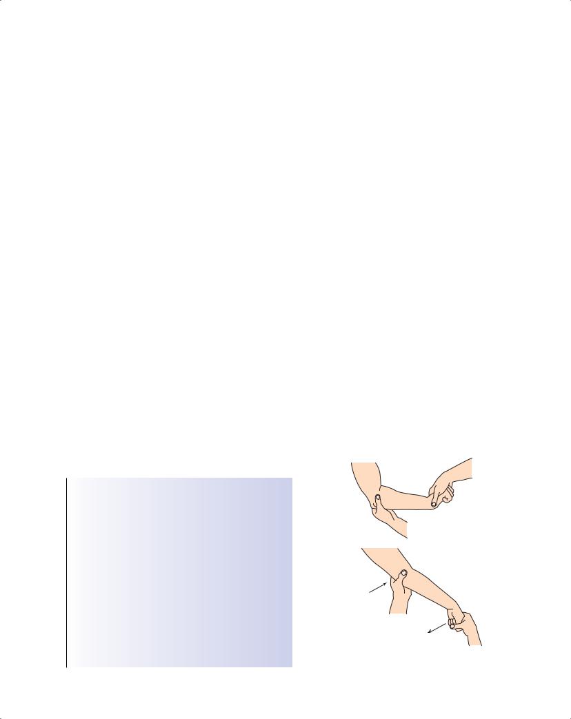
Both conditions are characterized by pain and tenderness over the respective epicondyle, and pain on resisted movements: resisted dorsiflexion of the wrist, middle finger, or both, in lateral epicondylitis (Figure 3.8), and resisted flexion of the wrist in medial epicondylitis. Both may be aggravated by repetitive movements and lifting. There may be night pain, early morning stiffness and stiffness after periods of inactivity. Pain referred from the neck or shoulder is distinguishable by less localized symptoms, associated neurological symptoms and the lack of local signs. Pain arising
Box 3.4 Differential diagnosis of elbow pain
•Lateral epicondylitis (“tennis elbow” or lateral elbow pain)
•Medial epicondylitis (“golfer’s elbow” or medial elbow pain)
•Olecranon bursitis
•Elbow joint
Rheumatoid arthritis
Septic arthritis
Osteoarthritis (rare)
•Cervical radiculopathy
•Tendinopathies
Biceps tendinopathy (anterior elbow pain)
Triceps tendinopathy, avulsion (posterior elbow pain)
•Nerve compression, entrapment
Ulnar neuropathy
Median nerve
Anterior interosseous nerve
Cubital tunnel syndrome
from the elbow joint is usually more posterior and less well localized and may be associated with an elbow effusion and difficulty straightening the elbow because of restriction.
Lateral and medial epicondylitis are generally self-limiting and patients should be informed of the generally favourable prognosis. In a general practice trial, 80% of patients with elbow pain of already greater than 4 weeks’ duration were recovered after 1 year, simply following an expectant policy without any specific treatment. Prognostic factors found to be at least moderately associated with a poorer outcome at 1 year include previous occurrence, high physical strain at work, manual jobs, high baseline levels of pain and/or distress, passive coping and less social support.
Treatment—Interventions have mainly been tested for lateral epicondylitis, but the results are probably generalizable to medial epicondylitis. Treatment in the acute stage involves relative rest and avoidance of specific activities that aggravate the discomfort. Ice may be applied, but there are no data about its effects. Use of a tennis elbow brace or strap is common and may provide shortterm pain relief while worn, allowing some return to activity.
Topical and oral NSAIDs may provide short-term relief of pain, although evidence is limited. Local skin reactions may occur with topical treatment.
Stretching and strengthening exercises may be helpful. Most studies have assessed their effect as part of multimodal interventions involving mobilization techniques at the elbow other physical therapies, with mixed results.
Corticosteroid injection with local anaesthetic may provide short-term pain relief (less than 3 months), although over the long term may be less effective than no treatment or physiotherapy (consisting of ultrasound, deep friction massage and an exercise programme). After an initial favourable response lasting 6 or more weeks, there may be a recurrence of symptoms. It is important to consider the close proximity of the ulnar nerve when performing
Patient |
Examiner |
|
Stabilise
Extension
Flexion
Figure 3.8 Provocation tests for lateral epicondylitis. Resisted extension of
the middle finger also elicits pain
20 ABC of Rheumatology
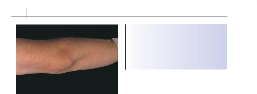
Figure 3.9 Olecranon bursitis—e.g. tophaceous gout at the elbow
steroid injection for medial epicondylitis. Adverse effects of injection are generally mild and transient and include post-injection pain, depigmentation and local skin and subcutaneous atrophy.
Trials of ultrasound have been conflicting but have generally reported marginal or no benefit, while laser therapy trials and trials of various other physical therapies have consistently been negative. There is no strong and consistent evidence that ESWT provides benefits in terms of pain and function in lateral epicondylitis. Acupuncture (needle, laser or electro-acupuncture) may provide short-term pain relief. Botulinum toxin injection and topical glyceryl trinitrate have recently been proposed as treatments for lateral epicondylitis, but further research is required before these therapies can be recommended.
Surgery is reserved for patients with recalcitrant, limiting symptoms, although evidence of benefit from controlled trials is limited. The most common operations are open excision, debridement and release and/or repair of the extensor or flexor tendon origins at the lateral or medical epicondyle. Percutaneous and arthroscopic procedures have also been described.
Other elbow disorders
Arthritis of the elbow joint may be due to systemic inflammatory arthritides, including rheumatoid and seronegative arthritis, crys- tal-induced synovitis (gout or pseudogout) and, rarely, septic arthritis. Neisseria gonococcus arthritis should be suspected in atrisk individuals. Osteoarthritis of the elbow is rare and usually relates to prior fractures or trauma.
Olecranon bursitis (“student’s elbow”) presents with discrete swelling, pain and inflammation at the posterior point of the elbow and may be caused by acute or repetitive trauma, crystals or sepsis (Figure 3.9, Box 3.5). The presence of nodules suggests either rheumatoid arthritis (seen in active disease or as a side effect of methotrexate) or gout (tophi). Infection may follow an abrasion or initial cellulitis and the most common causative organism is Staphylococcus aureus. Systemic symptoms such as fever, leucocytosis and elevated inflammatory markers occur with sepsis and
Box 3.5 Causes of olecranon bursitis
•Trauma (acute or chronic)
•Sepsis
•Metabolic or crystals
•Inflammatory arthritis
•Uraemia
•Calcific deposits
•Idiopathic
crystals. When olecranon bursitis is suspected, blood cultures and aspiration for crystals, Gram stain, and culture are essential.
Steroid injection is often helpful for olecranon bursitis due to inflammatory or crystal arthritis. Broad-spectrum antibiotics and possibly open drainage and lavage are used when there is sepsis.
Entrapment or inflammation of the ulnar, radial and median nerves can cause neurological disturbances involving the elbow and forearm. Paraesthesia and numbness involving the fourth and fifth fingers accompanied by weakness of the interossei may be caused by ulnar neuropathy, the most common compression neuropathy affecting the elbow. Tapping over the ulnar groove (Tinel’s sign) may reproduce pain or numbness in the fourth and fifth fingers. Nerve conduction studies are helpful in diagnosis. Management depends on the severity and cause.
Reference
Speed C, Hazleman B, Dalton S. Fast Facts: Soft Tissue Rheumatology. Health Press, Oxford, UK, 2000.
Further reading
Arthritis Research Campaign. The Painful Shoulder. An information booklet. Arthritis Research Campaign, York, UK, 2009. Available online at http:// www.arc.org.uk/arthinfo/patpubs/6039/6039.asp
Binder A. Neck pain. Clinical Evidence 2006; 15: 1654–1675.
Bjelle A. Epidemiology of shoulder problems. Baillière’s Clinical Rheumatology 1989; 3: 437–451.
Buchbinder R, Green S, Struijs P. Tennis elbow. Clinical Evidence 2008. Available online at http://clinicalevidence.bmj.com
Dalton S. Clinical examination of the painful shoulder. Baillière’s Clin Rheumatol 1989; 3: 453–474.
Hadler NM. Coping with arm pain in the workplace. Clinical Orthopaedics and Related Research 1998; 351: 57–62.
McClune T, Burton AK, Waddell G. Whiplash associated disorders: a review of the literature to guide patient information and advice. Emergency Medicine Journal 2002; 19: 499–506.
Mitchell C, Adebajo AO, Hay E, Carr A. Shoulder pain: diagnosis and management in primary care. British Medical Journal 2005; 331: 1124–1128.
Royal College of Radiologists. Making the Best Use of a Department of Clinical Radiology: Guidelines for Doctors. Royal College of Radiologists, London, 1999.
Speed C. Shoulder pain. Clinical Evidence 2005; 14: 1543–1560.

CHAPTER 4
Low Back Pain
Rajiv K Dixit1 and D John Dickson2
1University of California, San Francisco, USA
2Redcar & Cleveland Primary Care Trust, Redcar, UK
OVERVIEW
•Most patients with low back pain improve within 4 weeks.
•Degenerative change in the lumbar spine is the most common cause of pain.
•Imaging studies are rarely indicated, unless symptoms persist beyond 4 weeks.
•Imaging abnormalities should be carefully interpreted as they are frequently present in asymptomatic people.
•Most patients respond to a programme that includes analgesia, education, back exercises, aerobic conditioning and weight control. Surgery is rarely needed.
Low back pain (LBP) is the most common musculoskeletal symptom and poses a major socio-economic burden. An estimated 80% of the population will experience back pain during their lifetime; 90% of these patients will have resolution of their symptoms within 4 weeks.
Sciatica is the result of nerve root impingement and occurs in <1% of patients. The pain is radicular (and almost invariably radiates below the level of the knee) in the distribution of a lumbosacral nerve root, sometimes accompanied by sensory and motor deficits. Sciatica should be differentiated from non-neurogenic sclerotomal pain, which arises from pathology within the disc, facet joint or paraspinal muscles and ligaments. Sclerotomal pain is nondermatomal in distribution and often radiates into the lower extremities but not below the knee or with associated paraesthesiae as with sciatica.
Causes of LBP
LBP usually originates from the lumbar spine (Figure 4.1); pain is rarely referred to the spine from other structures (Box 4.1). Over 95% of LBP is mechanical. Mechanical pain is generally due to an anatomical abnormality that increases with physical activity and is relieved by rest and recumbency. Systemic disease (infection, neoplasm and spondyloarthropathy) accounts for only 1–2% of LBP.
ABC of Rheumatology, 4th edn. Edited by Ade Adebajo. ©2010 Blackwell Publishing Ltd. 9781405170680.
Box 4.1 Causes of LBP
Mechanical
•Lumbar spondylosis*
•Disc herniation*
•Spondylolisthesis*
•Spinal stenosis*
•Fractures (mostly osteoporotic)
•Idiopathic (“non-specific”)
Neoplastic
•Primary
•Metastatic
Inflammatory
• Spondyloarthropathies
Infectious
•Vertebral osteomyelitis
•Epidural abscess
•Septic discitis
Metabolic
•Osteoporotic compression fractures
•Paget’s disease
Referred pain to spine
•From major viscera, retroperitoneal structures, urogenital system, aorta, or hip
*Related to degenerative changes
Lumbar spondylosis
The most common cause of mechanical LBP is degenerative change. In lumbar spondylosis (lumbar osteoarthritis) degenerative changes occur in the intervertebral disc and facet joint. Imaging evidence of lumbar spondylosis (disc space and facet-joint narrowing, osteophytes and subchondral sclerosis) is common, increases with age and is often asymptomatic.
Disc herniation
The nucleus pulposus in a degenerated disc may prolapse and push out the weakened annulus, usually posterolaterally. Imaging evidence of disc herniation is common even in asymptomatic adults. Occasionally, disc herniation may result in nerve root impingement (Figure 4.2), causing sciatica. Of all clinically significant
21