Материал: ABC-of-Rheumatology-ABC-Series-
Pain in the Wrist and Hand |
7 |
|
|
|
|
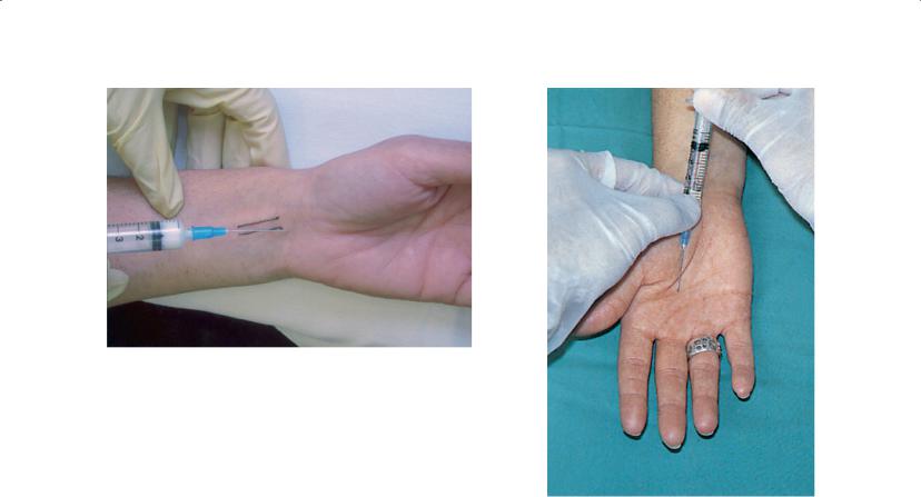
Figure 2.3 Injection technique for carpal tunnel syndrome
steroid preparation, e.g. hydrocortisone acetate, often precipitates the symptoms, but it is effective and non-toxic (O’Gradaigh and Merry, 2000; Wong et al., 2001).
Recurrent daytime symptoms, unrelieved by splints, warrant nerve-conduction studies. Slowing of median nerve conduction at the wrist suggests demyelination due to local compression. The action potential is reduced or absent due to nerve-fibre loss if the lesion is severe or prolonged. Needle electromyography is unpleasant but detects denervation.
Decompression surgery should be considered for: recurrent symptoms not eased by splints or injection; significant nerve damage; muscle wasting; and/or permanent numbness (Trumble et al., 2001). Pins and needles often worsen briefly post-operatively while the nerve recovers. Recovery of sensation or strength, or both, may be limited or non-existent if the lesion is severe and longstanding.
Finger flexor tendonosis and trigger finger
Gripping and hard manual work cause palpable thickening and nodularity of the finger flexor tendon; tendon sheath synovitis may also be present. The affected fingers are stiff in the morning, when the patient also has pain in the palm and along the dorsum of the finger(s). The pain is reproduced by passive extension of the finger. This is common in rheumatoid arthritis and in dactylitis caused by seronegative arthritis. Nodular flexor tenosynovitis is more common and less responsive to treatment in patients with diabetes than in other patients (Stahl et al., 1997).
Trigger finger is caused by a nodule catching at the pulley that overlies the metacarpophalangeal joint in the palm. The patient wakens with the finger flexed and has to force it straight with a painful or painless click. Triggering also occurs after gripping. The nodule and the “catch” in movement are felt in the palm.
Management and injection technique—A low-pressure injection of local anaesthetic followed by a locally acting steroid preparation alongside the tendon nodule in the palm helps (Rankin and
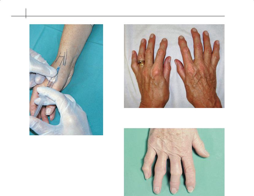
Figure 2.4 Injection technique for flexor tenosynovitis and trigger finger
Rankin, 1998) (Figure 2.4). If symptoms are persistent or recurrent, surgical release is needed.
Overuse and local injury (after opening a tight jar) are the most common causes of thumb flexor tenosynovitis and trigger thumb. Either the interphalangeal joint cannot be flexed or it sticks in flexion and snaps straight. The sesamoid bone in the flexor pollicis brevis tendon is tender on the volar surface of the thumb’s metacarpophalangeal joint. Corticosteroid injection next to the sesamoid bone at the site of maximal tenderness helps.
De Quervain’s tenosynovitis
De Quervain’s stenosing tenosynovitis affects the tendon sheath of abductor pollicis longus and extensor pollicis brevis at the radial styloid. It causes pain at or just proximal or distal to the styloid, in contrast with first carpometacarpal osteoarthritis, which causes pain at the base of the thumb. Tenderness, swelling and Finkelstein’s test—pushing the thumb into the palm while holding the wrist in ulnar deviation—increases the pain. Crepitus or a tendon nodule may cause triggering.
Management and injection technique—Rest is essential, with avoidance of thumb extension and pinching, but immobilization splints are inconvenient. Therapeutic ultrasound or local anti-inflamma- tory gels help; injection of local anaesthetic, then a locally acting steroid preparation alongside the tendon under low pressure at the point of maximum tenderness rapidly relieves the pain (Figure 2.5). A second injection may be needed. Surgery is rarely necessary, unless stenosis or nodule formation develops.
8 ABC of Rheumatology
Figure 2.6 Nodal osteoarthritis (Heberden’s nodes) and first carpometacarpal osteoarthritis
Figure 2.5 Injection technique for de Quervain’s tenosynovitis
Extensor tenosynovitis
Inflammation of the common extensor (fourth) compartment causes well-defined swelling that extends from the back of the hand to just proximal to the wrist. The extensor retinaculum causes a typical “hourglass” shape proximal and distal to the wrist. This contrasts with wrist synovitis, which causes diffuse swelling distal to the radius and ulna. Repetitive wrist and finger movements, especially with the wrist in dorsiflexion, are the cause, and this is one of the several causes of forearm and wrist pain seen in keyboard workers. It is also common in rheumatoid arthritis. Rest helps extensor tenosynovitis, but often a corticosteroid injection into the tendon sheath is needed. Workplace reviews and wrist supports for those who use a keyboard and mouse help prevent recurrences.
Mallet finger
This is a flexion deformity affecting the distal interphalangeal joint of the finger and is due to either distal extensor tendon rupture or avulsion with a bony fragment after traumatic forced flexion of the extended fingertip. The resultant weakness is often painless and presents with an inability to actively extend the fingertip. Treatment is usually by splinting the distal interphalangeal joint in extension or, rarely, surgery.
Osteoarthritis
Nodal osteoarthritis
Nodal osteoarthritis most commonly involves the distal interphalangeal joints and is familial. The joint swells and becomes inflamed and painful, but the pain subsides over a few weeks or
Figure 2.7 Nodal osteoarthritis with Bouchard’s nodes
months and leaves bony swellings (Heberden’s nodes). Most patients manage with local anti-inflammatory gels or no treatment once they know the prognosis is good. The appearance sometimes causes distress. Occasionally, the joint becomes unstable and limits pinch gripping. Surgical fusion of the index distal interphalangeal joints or thumb interphalangeal joint in slight flexion improves grip, although this is rarely necessary. Involvement of the proximal interphalangeal joints (Bouchard’s nodes) is less common and may be mistaken for early rheumatoid arthritis (Figure 2.6). Stiffness of the proximal joints impairs hand function significantly.
First carpometacarpal osteoarthritis
Pain at the base of the thumb in the early phase of first carpometacarpal osteoarthritis (Figure 2.7) is disabling, but with time the joint stiffens and adducts, and pain and disability decrease. The hand becomes “squared”. Management is usually conservative, but
Pain in the Wrist and Hand |
9 |
|
|
|
|
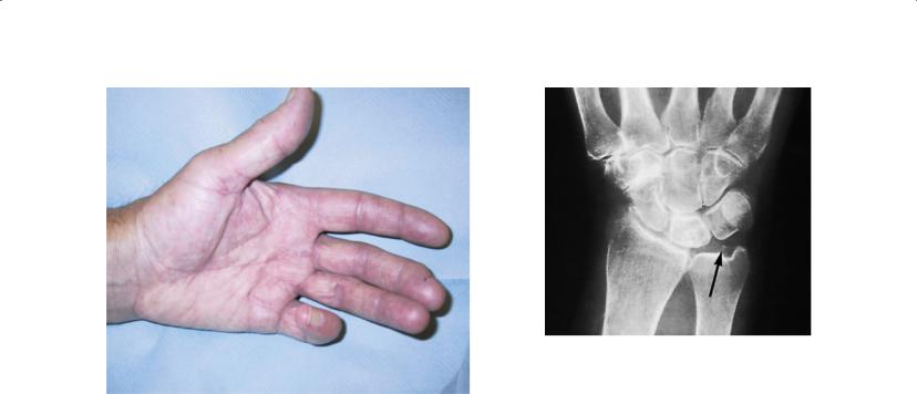
Figure 2.8 Dupuytren’s contracture
a corticosteroid injection helps severe pain associated with local inflammation. Surgical replacement is rarely warranted, although the outcome is good. Some find a splint helpful.
Dupuytren’s contracture
Dupuytren’s contracture (Figure 2.8) is a relatively common and painless condition that is associated with palpable fibrosis of the palmar aponeurosis, usually in the palm but occasionally at the base of a digit. It is more common in white people, men, heavy drinkers, smokers and patients with diabetes mellitus. The cause is unknown, but repeated trauma may be important. Fibroblast proliferation starts in the superficial fascia and invades the dermis. An early sign is skin pitting or puckering. The contraction eventually causes flexion of the digit(s), most often the ring finger, but disability is often minimal. Disabling and progressive flexion is more common in the familial form. Nodular fibromatosis also affects the sole of the foot, the knuckle pads (Garrod’s pads) and the penis (Peyronie’s disease), and these conditions may coexist. Specialist hand clinics use magnetic resonance imaging to assess the lesion. The role of local corticosteroid injections and radiotherapy in early disease is unclear (Ketchum and Donahue, 2000; Seegenschmiedt et al., 2001). Surgical excision is helpful but recurrence is common. No controlled studies exist.
Cubital tunnel syndrome
Ulnar nerve compression at the elbow can be caused by direct pressure from leaning on the elbow, stretching the nerve with the elbow in prolonged flexion at night, or holding a telephone. It causes pins and needles in an ulnar distribution (little finger and the ulnar side of the ring finger). Prolonged entrapment causes hypothenar wasting and weakness of the hand’s intrinsic muscles. The nerve is tender and sensitive at the elbow, where Tinel’s sign is positive. Nerve conduction studies are normal in around 50% of cases. Avoidance of direct pressure and prolonged elbow flexion help.
Figure 2.9 Chondrocalcinosis in wrist
Surgical anterior transposition of the nerve is occasionally needed. In some cases the ulnar nerve is compressed in Guyon’s canal at the wrist.
Systemic disorders causing hand pain
Inflammatory arthritis
The hands are often affected early in rheumatoid arthritis, with symmetrical swelling of the metacarpophalangeal joints, proximal interphalangeal joints and wrists. The feet and other joints are usually also affected. Psoriatic and other forms of seronegative arthritis are less common, are more likely to be asymmetrical, and may be associated with marked skin and tendon changes that produce a “sausage” finger. The distal interphalangeal joints and adjacent nails may also be affected in psoriasis. Morning pain and stiffness are typical. Intra-articular steroids are often useful adjuncts to systemic medication.
Acute pseudogout and chondrocalcinosis of the wrist
Sudden wrist inflammation in an older patient may be due to calcium pyrophosphate arthritis (pseudogout). Marked swelling and inflammation are observed—the joint feels hot, and infection may need to be excluded. Chondrocalcinosis (Figure 2.9), although often asymptomatic, is usually seen in the triangular ligament of the wrist on X-ray radiography. The joint aspirate is turbid and contains weakly positively birefringent crystals under polarized light. Steroid injection or a short course of a non-steroidal antiinflammatory drug or colchine usually helps; regular use of nonsteroidal anti-inflammatory drugs or colchine can be used to manage frequent attacks.
Acute gout and chronic tophaceous gout
Acute urate gout rarely affects the hands. Tophaceous deposits in individuals in renal failure or who have been on long-term diuretic treatment are initially painless, chalky subcutaneous deposits. The tophi can ulcerate and a few such patients also develop acute gout in the hand and elsewhere.
10 ABC of Rheumatology
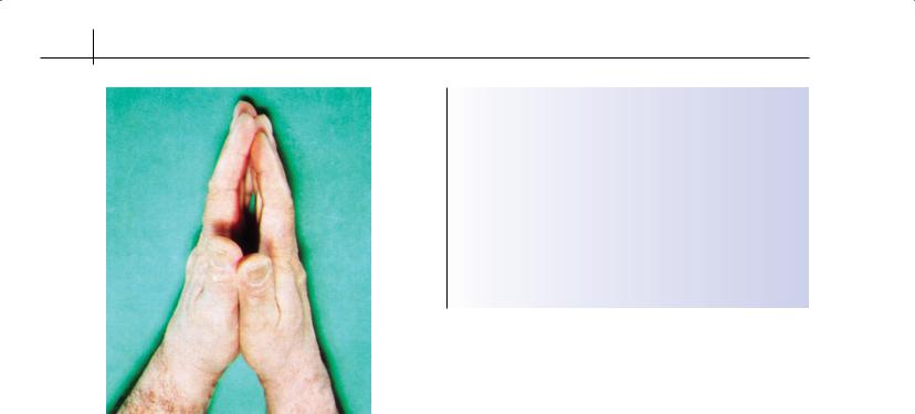
Figure 2.10 Positive prayer sign: diabetic stiff hands (also nodal osteoarthritis and flexor tenosynovitis)
Diabetic stiff hand (cheiroarthropathy—limited joint mobility syndrome)
Stiff hands are seen in 5–10% of patients with type I diabetes. This is more common in those with poor diabetic control and is associated with limited shoulder mobility, diabetic nephropathy and retinopathy. Patients develop waxy, tight skin and a so-called positive prayer sign—inability to hold the fingers and palms together (Figure 2.10). However, limited joint mobility in diabetes is multifactorial, and may also be due to flexor tenosynovitis, Dupuytren’s contracture or nodal osteoarthritis. Good diabetic control is essential. Injection for symptomatic flexor tenosynovitis helps. No specific treatment exists for the skin changes.
Raynaud’s phenomenon
This disorder, which results from severe vasospasm in response to a temperature change, causes marked and typically sharply demarcated pallor of one or more digits. As circulation recovers, the digit becomes blue (cyanotic) and then bright red because of rebound hyperaemia—the triphasic response. Raynaud’s is commoner in females than males. In young women the condition is often a harmless nuisance, requiring warm gloves and sometimes vasodilators. Its onset for the first time in older people warrants investigation. Raynaud’s may also be part of a systemic autoimmune disorder (rheumatoid arthritis, systemic lupus erythematosus, or systemic sclerosis), and it occasionally leads to necrosis. Autoimmuneassociated Raynaud’s can be extremely severe and requires specialist referral. Vibration white finger is a compensational industrial disease in people who use vibrating tools. Roughly two in three patients with primary Raynaud’s phenomenon have spontaneous resolution of their symptoms (Spencer-Green, 1998).
Box 2.1 Characteristics of chronic upper limb pain syndrome
•Often starts as carpal tunnel syndrome, flexor tenosynovitis or tennis elbow
•Spreads to affect the upper arm more diffusely
•Physical signs may be minimal
•Often associated with:
use of keyboards
sudden changes in work practices
disharmony at work
anxiety and sleeplessness
•Neurophysiological and psychosocial mechanisms involved
•Best dealt with non-judgementally
Other disorders
Ganglion
A ganglion is a cystic swelling in continuity with a joint or tendon sheath through a fault in the capsule. It is filled with clear, viscous fluid rich in hyaluronan. Ganglia are common on the dorsal wrist, are often painless and resolve spontaneously (50% at 6 years; see http://www.medicine.ox.ac.uk/bandolier/booth/miscellaneous/ wristgang.html). Often, only reassurance of the patient is required. Wrist splints relieve the pain. Aspiration and injection are rarely effective, and surgical excision is best if the ganglion is persistent and painful.
Chronic (work-related) upper limb pain
The main symptom of chronic upper limb pain is pain (Box 2.1). A local cause (carpal tunnel syndrome, flexor or extensor tenosynovitis, or tennis elbow) may be the initial trigger. The patient develops widespread pain that is often disproportionate to the findings but causes great distress. A prior change in work pattern may exist, and often disharmony is found at the workplace. The cause is unclear, but neurophysiological and psychosocial factors are probably involved. The phenomenon of central “wind up” of pain seen in many chronic pain syndromes probably plays a role. It is easy for the doctor to find the problem exasperating and difficult to understand, but it is best managed non-judgementally. Early reductions in work activities and pain-control measures are important, but it is best not to ask the person to take too much time off. Advice to the employer to review work practices reduces the risk of litigation. Referral to a specialist pain clinic should be considered.
Osteonecrosis (rare)
Kienböck’s disease is the late result of a dorsiflexion injury often seen in manual labourers. Fragmentation and collapse of the lunate causes shortening of the carpus and secondary osteoarthritis. Osteonecrosis takes up to 18 months to appear on X-ray radiography.
Scaphoid bone fracture
Pain in the anatomical snuffbox after a fall onto an outstretched hand requires an immediate X-ray examination, although a

Pain in the Wrist and Hand |
11 |
|
|
|
|
fracture is not always visible. Any severe wrist injury should be managed as a potential scaphoid fracture with a plaster, and a further X-ray radiograph should be taken 3 weeks later. Unrecognized scaphoid fracture leads to pain associated with failed union, osteonecrosis and secondary osteoarthritis.
Writer’s cramp
Writer’s cramp is the most common type of focal dystonia and occurs during complex hand activities—writing or playing a musical instrument. Clumsiness and painful tightness in the hand and forearm occur during writing or playing, and abnormal tension and strange posturing develop. Focal dystonias are often inappropriately described as “psychological.” Local botulinum toxin injection produces temporary relief. Retraining and learning new techniques help some patients, but the outlook is poor and may lead to the end of musical careers.
Septic arthritis
Septic arthritis of the hand or wrist is rare. It is an important differential diagnosis of acute pseudogout. If septic arthritis is suspected, it should be treated as a medical emergency and referred to Accident and Emergency or a specialist unit for investigation and appropriate intravenous antibiotic treatment. The patient is usually febrile and unwell. It is essential not to start antibiotics before all the necessary samples have been taken for culture. Non-steroidal anti-inflammatory drugs and analgesics can be given for pain, which is often severe.
Local corticosteroid injection technique
During |
local corticosteroid injections (Box 2.2), an injection |
of local |
anaesthetic (or topical anaesthetic) is followed by |
0.2–1 ml of a suitable steroid preparation, such as hydrocortisone acetate 25 mg/ml or depot methylprednisolone 40 mg/ml. Methylprednisolone is about five times as powerful as hydrocortisone on a mg per mg basis. It is best first to introduce the needle
Box 2.2 Local corticosteroid injection technique
•Hand and arm well supported
•Equipment readily to hand
•Clean skin thoroughly
•Use small-bore needle
•Inject small volume of local anaesthetic
•Inject corticosteroid through same needle
•Always inject under low pressure
with local anaesthetic and then to inject the steroid under low pressure. Patients should be warned that the pain might increase for a day or two after injection. Superficial injections or, very rarely, leakage of the corticosteroid along the needle track, cause local skin depigmentation and atrophy of subcutaneous fat; this is more likely with depot injections of steroid. Consent from the patient should always be obtained.
References
Kamath V, Stothard J. Erratum to: A clinical questionnaire for the diagnosis of carpal tunnel syndrome. Journal of Hand Surgery (Edinburgh, Scotland)
2004; 29: 95.
Ketchum LD, Donahue TK. The injection of nodules of Dupuytren’s disease with triamcinolone acetonide. Journal of Hand Surgery 2000; 25: 1157–1162.
O’Gradaigh D, Merry P. Corticosteroid injection for the treatment of carpal tunnel syndrome. Annals of the Rheumatic Diseases 2000; 59: 918–919.
Pal B, O’Gradaigh D, Merry P. Diagnosis of carpal tunnel syndrome. Rheumatology 2001; 40: 595–597.
Rankin ME, Rankin EA. Injection therapy for management of stenosing tenosynovitis (de Quervain’s disease) of the wrist. Journal of the National
Medical Association 1998; 90: 474–476.
Seegenschmiedt MH, Olschewski T, Guntrum F. Radiotherapy optimization in early-stage Dupuytren’s contracture; results of a randomized study.
International Journal of Radiation Oncology, Biology, Physics 2001; 49: 785–798.
Spencer-Green G. Outcomes in primary Raynaud’s phenomenon: a metaanalysis of the frequency, rates and predictors of transition to secondary diseases. Archives of Internal Medicine 1998; 158; 595–600.
Stahl S, Kanter Y, Karnelli E. Outcome of trigger finger treatment in diabetes.
Journal of Rheumatology 1997; 24: 931–936.
Trumble TE, Gilbert M, McCallister WV. Endoscopic versus open surgical treatment of carpal tunnel syndrome. Neurosurgery Clinics of North America 2001; 12: 255–266.
Wong SM, Hui ACF, O’Gradaigh D, Merry P. Corticosteroid injection for the treatment of carpal tunnel syndrome. Annals of the Rheumatic Diseases 2001; 60: 897.
Yagev Y, Carel RS, Yagev R. Assessment of work-related risk factors for carpal tunnel syndrome. Israel Medical Association Journal 2001; 3: 569–571.
Further reading
Bland JDP. Carpal tunnel syndrome. British Medical Journal 2007; 335: 343–346.
Zhang W, Doherty M, Leeb BF et al. EULAR evidence based recommendations for the management of hand osteoarthritis: report of a Task Force of the EULAR Standing Committee for International Clinical Studies Including Therapeutics (ESCISIT). Annals of the Rheumatic Diseases 2007; 66: 377–388.