Материал: ABC-of-Rheumatology-ABC-Series-

2 ABC of Rheumatology
patients to self care has been shown to reduce their GP visits by 40–69% (Schillinger et al., 2003). Patient education is not a treatment in itself but a treatment enhancer, magnifying the effects of standard treatments by persuading patients to adhere to them more closely, or to adopt actions that are believed to be beneficial. To do this, patients must be active collaborators in their care and believe in their ability to perform a specific task or achieve a certain objective. This is known as “self-efficacy”. For changes to occur patients must acquire knowledge and skills, and so patient education involves the multidisciplinary team and the patient and their partner/carers in both primary and secondary care. Every consultation is an opportunity to educate and provide information. In order to care for themselves, patients will need to know about the topics shown in Box 1.2.
Patients should be given both verbal and written explanations. The Arthritis Research Campaign (arc), Arthritis Care, and the National Rheumatoid Arthritis Society are good reliable sources of the latter.
Skill enhancement can be gained from attendance at an Expert Patients Programme, and giving the patient the address of local and national community support networks offers great benefits.
It is important to remember that simply because information has been provided does not mean that it has been understood or acted upon. One quick and easy method to ensure assimilation is the “teach me back” method (Schillinger et al., 2003), which involves the patient being asked to “teach me” or “show me” as if the professional does not understand the problem. This quickly identifies any misunderstandings and allows purposeful correction.
Box 1.1 Role of the specialist nurse
•Supervise treatment safety—e.g. monitoring disease-modifying antirheumatic drugs (DMARDS)
•Review treatment effectiveness
•Coordinate the multidisciplinary team
•Provide a communication channel between the patient and the team
•Act as the patient’s advocate
•Promote continuity of care
•Identify and address psychosocial patients’ issues
•Man telephone advice lines
•Facilitate education for patients, carers and health professionals
Box 1.2 Knowledge necessary for self care
•Disease aetiology and progress
•Drugs and how to take them; what the side effects are and what to do if they occur
•How to exercise
•How to protect joints and acquire appropriate devices and home changes
•How to control pain
•Coping strategies
Who should be referred to secondary care?
Waiting times for new rheumatology appointments vary widely and depend on local resources but also, to some extent, on how clinicians triage referrals from GPs. To make the system work effectively, care pathways need to be developed in which the patient is a partner, and which take psychosocial as well as biomechanical factors into consideration. The outcome, in terms of whether the patient is given an appropriate priority with an appropriate heathcare professional, depends largely upon the information contained within the referral letter. Standardized referral forms may help but have the disadvantages that they are time-consuming to complete and rather impersonal. Helpful information to include in a referral letter is shown in Box 1.3.
It has been estimated that 15–30% of all GP consultations are for musculoskeletal conditions. Most of these are for osteoarthritis in the over 50s age group and back pain in the under 50s. One challenge for the GP is how to spot the small number of patients with early inflammatory arthritis among this caseload who will benefit from early referral to hospital and prompt treatment with DMARDs. There are no specific clinical, radiological or immunological markers for rheumatoid arthritis (RA). Normal blood test results and X-rays do not exclude RA, but equally a positive rheumatoid factor does not clinch the diagnosis. Most rheumatology departments encourage an “inclusive approach” to referral and encourage GPs to maintain a high index of suspicion and not delay patients with possible inflammatory arthritis. Ideally, patients suspected of having inflammatory problems will be fast-tracked to secondary care. Box 1.4 highlights certain features thought to be indicative of early RA.
Box 1.3 Important information to include in a rheumatology referral letter
•Length of history
•Pattern of joint involvement
•Presence of joint swelling
•Presence of early morning stiffness
•Previous treatments and response
•Level of distress/disability
•Results of investigations
•Other relevant medical or psychosocial factors
Box 1.4 Symptoms and signs suggestive of early inflammatory arthritis
•Symmetrical soft-tissue swelling (synovitis) of wrists and/or metacarpophalangeal joints and/or proximal interphalangeal joints
•Joint stiffness a significant problem—especially in the early mornings for >30 minutes
•Soft-tissue swelling of any joints
•Good response to a trial of non-steroidal anti-inflammatory drugs

Community Rheumatology |
3 |
|
|
|
|
Primary care management of musculoskeletal problems
Clearly, the majority of patients presenting to GPs will not have inflammatory arthritis. Indeed, often a precise pathological diagnosis based on symptoms and signs and results of investigations will not be possible, and may not be the most appropriate approach to management. This “medical model” of care often fails to address other important influences on pain perception, such as emotional and behavioural factors, and may encourage chronicity by using terms such as “arthritis”, “wear and tear” or “degeneration”, which emphasise the unchanging nature of the condition. Doctors are trained to diagnose “disease”, whereas the patient’s concern is what to do about their musculoskeletal pain, not just what to call it.
An alternative approach, which may be more useful in primary care, limits the diagnostic process to identifying potentially serious pathology—the so-called “red flag” disorders—and other specific diseases or disorders. This system was initially developed for back pain, and has been effective in changing the primary care management of this condition. It is equally applicable to other widespread or regional pain disorders, however (Box 1.5) (reviewed in Carr, 2001). Patients with “red flags” and certain other patients with specific diagnoses, including inflammatory arthropathies and connective-tissue disorders, should be considered for referral to secondary care for further investigation and management.
Having excluded and dealt with the small proportion of patients with potential serious pathology and specific diagnoses, the next step is to decide how best to manage the remainder. Two areas need to be addressed: how to deal with the presenting pain and distress (discussed below), and how to prevent future disability. Guidelines for the management of low back pain highlight the importance of identifying factors that predict chronicity. It is important to give positive messages about likely recovery and lack of long-term
Box 1.5 “Red flags” for regional pain syndromes
History of significant trauma
•Fracture
•Major soft-tissue injury
Localized joint swelling and/or redness
•Septic arthritis
•Inflammatory arthritis
•Haemarthrosis
Unremitting night pain
•Malignancy
•Inflammation/infection
Bone tenderness
•Fracture
•Malignancy
•Infection
Systemic disturbance
Significant co-morbidity
harm, taking particular account of psychosocial barriers to recovery (“yellow flags”). These principles have been described elsewhere (Department of Health, 2007) and are summarized in Box 1.6.
Evidence-based primary care treatments for musculoskeletal problems
The shift in emphasis towards self-management of musculoskeletal problems means that the primary health-care team is of central importance. There is a growing evidence base supporting the effectiveness of a number of simple primary care interventions for musculoskeletal problems (reviewed in Schillinger et al., 2003). Direct access physiotherapy reduces wait times and costs for treatment and is one way to facilitate the use of exercise and self-management regimes. These have been demonstrated to be beneficial for patients with a variety of regional and widespread musculoskeletal conditions, including osteoarthritis, back pain, fibromyalgia and shoulder problems. Prescribed exercise need not be the province of the physiotherapist alone. Often, wait times to see a physiotherapist are excessively long, and many self-limiting musculoskeletal conditions can be managed with sensible exercise regimes undertaken outside the hospital setting. This has the advantage of promoting self-help and “demedicalizing” common musculoskeletal problems. arc publishes a wide range of patient information leaflets and booklets, which are useful adjuncts to advice and education provided by health-care professionals (Box 1.7).
Local steroid injections are effective for reducing pain from softtissue problems such as tennis elbow and shoulder problems in the short term but do not improve long-term outcome. They should be reserved for patients in whom pain is restricting rehabilitation
Box 1.6 Psychosocial factors that predict chronicity
•Belief that pain is due to progressive pathology
•Belief that pain represents harm or injury
•Belief that avoiding activity will speed up recovery
•Tendency to social isolation
•Tendency to anxiety/depression
•Expectation that passive treatments rather than self-help programmes will be of benefit
Box 1.7 arc publications
Arthritis Research Campaign (arc) leaflets, booklets and other publications are available from:
Dept RD
arc Trading Ltd Brunel Drive
Northern Road Industrial Estate Newark
Notts. NG24 2DE www.arc.org.uk

4 |
|
ABC of Rheumatology |
|
|
|
|
|
|
|
|
|
||
|
|
|
|
|
|
|
|
Box 1.8 Contraindications to local steroid injections |
|
Box 1.9 Evidence-based summary |
|||
|
|
|||||
|
Absolute |
|
• |
Early aggressive treatment of rheumatoid arthritis with disease- |
||
|
• |
Suspected septic arthritis |
|
|
modifying drugs improves clinical outcome and slows |
|
|
• |
Local skin disorders or sepsis |
|
|
radiological progression (O’Dell, 2001) |
|
|
• |
Active systemic infection |
|
• |
Psychosocial factors are the main predictors of chronic pain |
|
|
• |
Prosthetic joint |
|
|
disability (Linton, 2000) |
|
|
Relative |
|
• |
Local steroid injections provide effective short-term relief for |
||
|
|
|
soft-tissue problems but do not improve long-term outcome |
|||
|
• |
Poorly controlled diabetes mellitus |
|
|
||
|
|
|
(Smidt et al., 2002) |
|||
|
• Osteoporosis |
|
|
|||
|
|
|
|
|||
|
|
|
|
|
|
|
with the measures discussed above. Although the risks from local steroid injections are minimal, certain precautions need to be adhered to (Box 1.8).
Non-steroidal anti-inflammatory drugs may be beneficial for the short-term treatment of osteoarthritis but have a worrying sideeffect profile in the patient group most likely to be prescribed them (elderly females). Simple analgesics are the preferred option where possible.
Global issues
The issues discussed in this chapter have global application, as the burden of illness from musculoskeletal conditions is high in both the developed world and developing countries alike, particularly with an ever-increasing elderly population worldwide. In developing countries, it is essential to involve local community leaders and community health workers in the management of patients with these conditions. Awareness of the importance of musculoskeletal conditions, in terms of morbidity but also mortality, needs to be raised among all health-care workers, governments and members of the public. With increasing travel and migration, knowledge of the global spectrum of musculoskeletal conditions is important. There also needs to be an increasing emphasis on prevention through encouraging healthy lifestyles and joint protection and by tackling modifiable risk factors such as falls prevention. Whether in primary or secondary care, or whether in a developing or developed country, what is key is not where musculoskeletal care takes place, but that it is appropriately given.
Conclusion
Over the last 10 years there has been a shift in thinking about how best to care for patients with rheumatological disorders (Box 1.9). For those with inflammatory arthritis the emphasis is on prompt referral to secondary care so that treatment with potentially dis- ease-modifying agents can be instituted early, before irreversible joint damage has occurred. For patients with non-inflammatory
conditions, such as osteoarthritis and regional or widespread musculoskeletal pain, optimal management depends on developing an efficient triage system that can identify those with “red flags” who will benefit from referral to secondary care for further investigation and management. The first-line management for the remainder should be by health-care professionals in primary care, using the strategies outlined above.
References
Carr A. Defining the Extended Clinical Role for Allied Health Professionals in Rheumatology. arc Conference Proceedings No. 12. Arthritis Research Campaign, Chesterfield, 2001.
Department of Health. Self care for people with long term conditions. Department of Health Long Term Conditions Team, 2006; available online at http://www.dh.gov.uk/en/Healthcare/Longtermconditions/DH_087281 Linton SJ. A review of psychological factors in back and neck pain. Spine 2000;
25: 1148–1156.
Meadows A, Sheehan NJ. Prescribing and injecting: the expanding role of the rheumatology nurse. Musculoskeletal Care 2005; 3: 176–178.
O’Dell JR. Combinations of conventional disease-modifying antirheumatic drugs. Rheumatic Disease Clinics of North America 2001; 27: 415–426.
Schillinger D, Piette J, Grumbach K et al. Closing the loop: physician communication with diabetic patients who have low health literacy. Archives of Internal Medicine 2003; 163: 83–90.
Smidt N, Assendelft WJJ, Windt van der DAWM et al. Corticosteroid injections for lateral epicondylitis: a systematic review. Pain 2002; 96: 23–40.
Further reading
Dziedzic K, Jordan J, Sim J et al. Treatment options for regional musculoskeletal pain: what is the evidence? In: Breivik H & Shipley M, eds. Pain: Best Practice and Research Compendium. Elsevier, Toronto, 2007: 183–197.
Doherty M, Dougados M, eds. Osteoarthritis: current treatment strategies.
Best Practice and Research. Clinical Rheumatology 2001; 15: 517–656. Main CJ, Williams A. ABC of psychological medicine: musculoskeletal pain.
BMJ 2002; 325: 534–537.
White C, Cooper RG. In Practice: Prescribing and Monitoring of DiseaseModifying Anti-Rheumatic Drugs (DMARDs) for Inflammatory Arthritis. Arthritis Research Campaign, Chesterfield, 2005.

CHAPTER 2
Pain in the Wrist and Hand
Michael Shipley1 and Elspeth Wise2
1University College London Hospitals, London, UK
2Belmont Surgery, Durham, UK
OVERVIEW
•Nodal osteoarthritis affecting the distal interphalangeal joints is very common and generally painful only for a few months.
•If a patient presents with swollen and painful joints in the hand, consider inflammatory arthritis as a diagnosis.
•The hand and wrist are common sites for overuse and injury. Remember to ask about precipitating factors, especially work/ occupation and hobbies.
•Carpal tunnel syndrome is a common peripheral nerve entrapment syndrome and has a classical presentation.
•Raynaud’s syndrome generally requires symptomatic treatment only, but consider secondary causes if seen in older people.
Hand or wrist pain and resultant impaired function are often the cause of great anxiety for patients. Hands, as prehensile organs, give us a great deal of information about the world in which we live. They are capable of performing incredibly fine and delicate movements and are essential for work, sport, hobbies and social interaction.
Functional anatomy
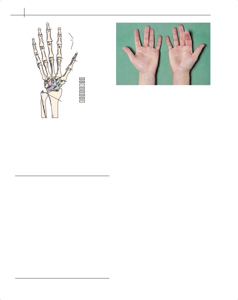
The wrist is a complex structure comprising three groups of joints: the radiocarpal joints, which allow flexion, extension, abduction, adduction and circumduction; the inferior radio-ulnar joint, which allows pronation and supination; and the intercarpal joints (Figure 2.1).
The eight carpal bones, in two rows of four, form a bony gutter and are the base of the carpal tunnel. The flexor retinaculum, a strong fascial band, forms the palmar side of the tunnel. Running through the carpal tunnel are the deep and superficial flexor tendons, the tendons of flexor pollicis longus, flexor carpi radialis and the median nerve. The ulnar nerve lies superficial to the flexor retinaculum but deep to the transverse carpal ligament in Guyon’s canal. The extensor tendons are held in position on the extensor surface of the wrist by the extensor retinaculum. Fibrous septa
ABC of Rheumatology, 4th edn. Edited by Ade Adebajo. ©2010 Blackwell Publishing Ltd. 9781405170680.
divide the extensor compartment into six. All of the flexor tendons are encased in a common synovial tendon, which extends from a position just proximal to the wrist to the middle of the palm. Flexor pollicis longus and flexor carpi ulnaris have their own individual sheaths, as do each of the six extensor compartments.
The hand bones are the metacarpals, proximal phalanges, middle phalanges, distal phalanges and sesamoid bones. A sesamoid bone lies at the base of the thumb in the tendons of flexor pollicis brevis. The first metacarpal bone of the thumb is the shortest and most mobile of the metacarpals and lies in a different plane to the others. This is important to allow opposition, i.e. pincer action to grasp objects. The carpometacarpal and trapezoscaphoid joints are prone to osteoarthritis.
Individual tendon sheaths for the deep and superficial flexor tendons start at the level of the distal transverse crease of the palm and end at the bases of the distal phalanxes. The sheath for flexor pollicis longus continues from the carpal tunnel to the distal phalanx. During flexion, five fibrous bands, or pulleys, hold the flexor sheaths in position.
The second to fifth metacarpophalangeal joints flex to about 90 °. Active extension is rarely more than 30 °. Passive extension varies from 60 ° to more than 100 ° in people with hypermobility. The proximal and distal interphalangeal joints are hinge joints. The lumbrical and interossei muscles produce complex movements that involve extension of the interphalangeal joints and flexion at the metacarpophalangeal joints and are essential to fine hand functions, such as writing.
There are many possible causes of pain in the wrist and hand (Table 2.1).
Tendon problems
Flexor tenosynovitis
Unaccustomed or repetitive use of the finger and inflammatory arthritis cause flexor tenosynovitis (Figure 2.2), inflammation of the synovial sheath of the finger flexor tendons, which leads to volar swelling and tenderness just proximal and distal to the wrist. The flexor tendon sheaths in the palm or finger may also be affected. The hand feels stiff, painful and swollen, particularly in the morning. Rest helps. Injection is sometimes needed. Local anaesthetic helps introduce the needle alongside the tendon in the palm just proximal to the metacarpophalangeal joint.
5
6 ABC of Rheumatology
DIPJ
Distal
|
PIPJ |
Middle Phalanges |
|
|
|
|
|
|
|
Proximal |
|
|
|
MCPJ |
|
|
|
IPJ |
|
Metacarpals |
|
MCPJ |
|
|
|
|
|
|
|
CMCJ |
Scaphoid |
|
|
Lunate |
|
Carpal bones |
|
|
Pisiform |
|
|
|
Triquetral |
|
Radius |
|
Hamate |
Ulna |
|
Capitate |
|
|
|
||
|
|
|
Trapezoid |
|
|
DRUJ |
Trapezium |
Figure 2.1 The bones of the hand. CMCJ = carpometacarpal joint; DIPJ = distal interphalangeal joint; DRUJ = distal radio-ulnar joint;
IPJ = interphalangeal joint; MCPJ = metacarpophalangeal joint; PIPJ = proximal interphalangeal joint
Table 2.1 Causes of pain in the wrist and hand
At all ages |
In older patients |
|||
|
|
|
|
|
• |
Trauma |
• |
Nodal osteoarthritis |
|
|
|
|
|
Distal interphalangeal joints |
|
|
|
|
(Heberden’s nodes) |
|
|
|
|
First carpometacarpal joints |
|
|
|
|
Proximal interphalangeal |
|
|
|
|
joints (Bouchard’s nodes) |
• |
Flexor tenosynovitis |
• |
Scaphoid fracture |
|
|
Carpal tunnel syndrome |
|
|
|
• Flexor tendonosis |
• Pseudogout |
|||
|
Trigger finger or thumb |
|
|
|
• De Quervain’s tenosynovitis |
• Gout |
|||
|
|
|
|
Acute |
|
|
|
|
Chronic tophaceous |
• |
Extensor tenosynovitis |
• Dupuytren’s contracture |
||
• |
Ganglion |
• |
Diabetic stiff hand |
|
• |
Mallet finger |
• |
Septic arthritis |
|
•Cubital tunnel syndrome
•Inflammatory arthritis
•Raynaud’s syndrome
•Writer’s cramp
•Chronic upper limb pain
•Reflex sympathetic dystrophy
•Scaphoid fracture
•Osteonecrosis
Figure 2.2 Flexor tenosynovitis
Carpal tunnel syndrome
Carpal tunnel syndrome is a peripheral nerve entrapment syndrome of the median nerve, often caused by flexor tenosynovitis. It can occur in the third trimester of pregnancy. Repetitive use of the hand increases the risk of developing carpal tunnel syndrome but its status as a work injury is controversial (Yagev et al., 2001). A ganglion, or very rarely amyloidosis or myxoedema, causes carpal tunnel syndrome. Pain, tingling and numbness in a median nerve distribution (thumb, index finger, middle and radial side of ring finger) are typically present on waking or can wake the patient. The fingers feel swollen and intense aching is felt in the forearm. The symptoms may appear when the patient holds a newspaper or the steering wheel of a car. Permanent numbness and wasting of the thenar eminence (flexor pollicis and opponens pollicis) cause clumsiness. The patient’s history often indicates the diagnosis (Pal et al., 2001), or using a scored questionnaire may help (Kamath and Stothard, 2004).
Tests and investigations—Tinel’s sign (tapping the median nerve in the carpal tunnel) or Phalen’s test (holding the wrist in forced dorsiflexion) may provoke symptoms. Weakness of abduction of the thumb distal phalanx with the thumb adducted towards the fifth digit is typical. The carpal tunnel and median nerve are seen on ultrasonic images, although US and MRI are not usually needed.
Management and injection technique—A splint worn on the wrist at night relieves or reduces the symptoms of carpal tunnel syndrome. This is diagnostic and may be curative. A corticosteroid injection into the carpal tunnel (Figure 2.3) may also be considered, as this often helps rapidly, although recurrence is common. The needle is inserted at the distal wrist skin crease, just to the ulnar side of the palmaris longus tendon, or about 0.5 cm to the ulnar side of flexor carpi radialis at an angle of 45 ° towards the middle finger. The local anaesthetic is injected superficially. If a small test injection of corticosteroid causes finger pain, the needle is in the nerve and needs to be repositioned. An injection of a locally acting