Материал: ABC-of-Rheumatology-ABC-Series-

CHAPTER 3
Pain in the Neck, Shoulder and Arm
Rachelle Buchbinder1 and Caroline Mitchell2
1Cabrini Hospital and Monash University, Melbourne, Australia
2University of Sheffield, Sheffield, UK
OVERVIEW
•Neck, shoulder and lateral elbow pain are common musculoskeletal problems for which patients seek care in general practice.
•Non-specific neck pain is often acute and self-limiting, attributed to a mechanical basis, but persistence or recurrence is common. For most patients with acute neck pain and no “red flags”, further investigation is not necessary.
•Neck pain usually responds to analgesia and advice about simple mobilization and exercises. High-quality evidence for the effectiveness of many treatment modalities is limited and often contradictory.
•Most shoulder complaints are due to rotator cuff disease, which is more prevalent with increasing age. Adhesive capsulitis, or “frozen shoulder”, is a self-limiting condition, occurring most commonly in middle age. It can be distinguished from rotator cuff disease by the presence of global restriction of shoulder movements. It is also more common in people with diabetes.
•For most patients with shoulder pain the diagnosis can also usually be made clinically. Treatment aims to control pain and restore movement and function of the shoulder.
•Lateral epicondylitis is thought to be an overload injury at the origin of the common extensors at the lateral epicondyle. Patients present with pain and tenderness over the lateral epicondyle and pain with resisted movements. Prognosis is generally favourable, with 80% recovery within a year. Management is directed towards controlling pain, avoiding aggravating activities and maintaining movement.
The neck and shoulder are two of the most common sources of musculoskeletal pain. Neck pain has a self-reported point prevalence of between 10 and 20%. The majority of neck pain is acute and self-limiting and can be attributed to a mechanical or postural basis. However moderate or severe symptoms may persist in up to 30% of patients.
Shoulder pain has a self-reported point prevalence of between 14 and 26% in the general population. The incidence of shoulder pain increases with age, as does its functional impact. About one-
ABC of Rheumatology, 4th edn. Edited by Ade Adebajo. ©2010 Blackwell Publishing Ltd. 9781405170680.
quarter of all new episodes presenting for care resolve fully within 1 month, and nearly half have resolved within 3 months of onset. However, persistence or recurrence of shoulder symptoms within a year of initial presentation is common (in up to 50% of people).
Anatomy and function of the neck and shoulder joint
The neck moves almost constantly during waking hours through flexion, extension and rotation at the intervertebral and facet joints of the seven cervical vertebrae, through the actions of the surrounding muscles.
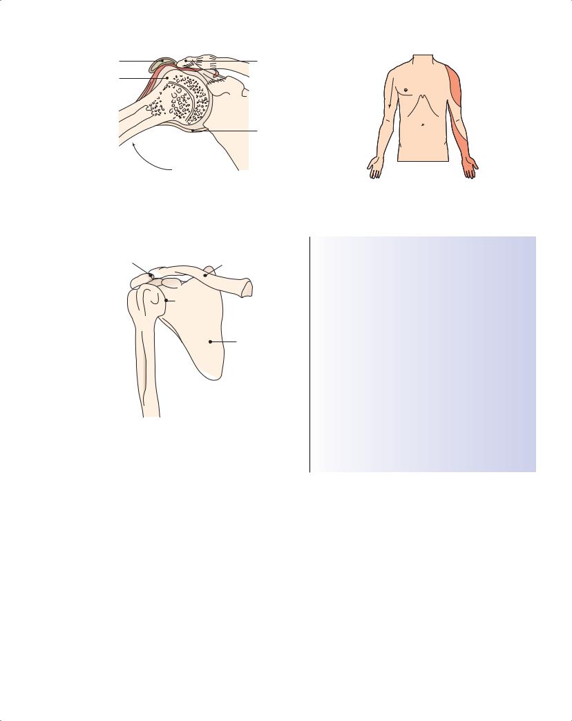
The shoulder is a series of articulations, including the scapulothoracic articulation, where the scapula slides on the ribcage (Figure 3.1). Soft tissue structures—capsules, ligaments, muscles, tendons, bursae and neurovascular elements—complete the framework and allow remarkable mobility to be achieved. The glenohumeral joint is extremely mobile and relies on the rotator cuff for stability. Instability, caused by laxity (congenital or acquired) or lack of muscular control because of pain, is a common feature of shoulder complaints.
The elbow is a compound synovial joint composed of a complex of two closely related articulations between the humerus and both the ulna and radius. It is supported by the ligaments and muscles.
Clinical evaluation
Neck and arm pain have a wide differential diagnosis. It is sometimes hard to distinguish between pain arising from the neck or the shoulder (Figure 3.2). Pain proximal to the shoulder, in the shoulder girdle or over the scapula indicates referred pain from the neck.
It is important to assess the patient’s concerns, expectations, functional disability and any psychosocial and occupational issues. Details of hand dominance, any injury, hobbies, sporting activities and treatments for this or any other similar previous musculoskeletal problems should be noted. Significant past and current medical history—prescribed drugs and adverse reactions—should also be explored. The history should elicit the presence of any clinical features that indicate potentially serious pathology.
Determine the mode of onset and duration of the pain, nature, site, radiation, temporal characteristics, exacerbating and relieving
12
Pain in the Neck, Shoulder and Arm |
13 |
|
|
|
|
Subacromial bursa |
Acromion |
Greater tuberosity
Rotator cuff 
Inferior joint capsule
Figure 3.1 The shoulder “complex” of joints. This includes the scapulothoracic articulation, where the scapula slides on the ribcage. Adapted from Speed et al., 2000
Acromioclavicular
joint |
Clavicle |
|
Subacromial 
space
Glenohumeral |
Subacromial joint |
|
joint |
||
|
||
|
Scapula |
Humerus 
Figure 3.2 Sites and radiation of pain in the shoulder, arm and neck. Adapted from Speed et al., 2000
Acromioclavicular joint 


 Sternoclavicular joint
Biceps 
 Glenohumeral joint/
  subacromial/rotator cuff/capsule
 Neck
Neck
Figure 3.3 Features of cervical-nerve-root lesions
Box 3.1 Differential diagnosis of neck pain
Structural
•Mechanical or non-specific
•Prolapsed intervertebral disc
•Cervical myelopathy
Neoplasm
• Primary or secondary
Inflammatory
•Rheumatoid arthritis
•Polymyalgia rheumatica and giant cell or temporal arteritis
•Spondyloarthropathies
Infection
•Discitis
•Osteomyelitis
•Paraspinal abscess
Metabolic
• Paget’s disease
Myofascial
• Myofascial syndromes, fibromyalgia
features, and associated symptoms. Disturbed sleep is common with both neck and shoulder pain. Nevertheless, nocturnal pain should raise suspicion of nerve root pain, bony pathology or underlying malignancy, particularly if there is a history of cancer and/or systemic symptoms.
Radiation of pain distally from the upper arm or elbow suggests referred pain from the neck or peripheral neurological lesions (Figure 3.2). Neurological symptoms should be sought and their distribution ascertained (Figure 3.3).
Other notable symptoms include stiffness, clicking, clunking or locking. Joint swelling around the shoulder or elbow can occur in relation to arthropathy, infection or trauma. Systemic symptoms, such as fevers, night sweats, weight loss, generalized joint pains, new “lumps” (lympadenopathy, mass lesions) and new respiratory symptoms, should be specifically sought.
A structured examination aims to define the source of the pain and the degree of functional deficit and coexisting pathologies.
It includes careful inspection, palpation, movement, special tests, neurological assessment and further investigations, as appropriate.
Neck pain
Pain in the neck usually arises because of poorly defined mechanical influences, although it can occur because of pathology within the spine or be referred from elsewhere. A list of differential diagnoses of neck pain is shown in Box 3.1. When considering the diagnosis it is important to look for “red flags” or clinical features that indicate that there might be a serious underlying cause of the complaint (Table 3.1). Restricted cervical movements and local tenderness help to confirm the local origin of neck pain. Risk

14 ABC of Rheumatology
Table 3.1 “Red flags” or clinical features indicative of potentially serious pathology in the neck and/or shoulder
“Red flags” |
Potential pathology |
|
|
History of cancer, symptoms and signs of |
Malignancy |
cancer, unexplained deformity, mass or |
|
swelling |
|
Fever, systemically unwell, redness and |
Infection |
swelling |
|
Trauma, epileptic fit, electric shock, loss of |
Unreduced shoulder |
rotation and normal shape |
dislocation |
Recent trauma, acute disabling pain and |
Acute rotator cuff tear |
significant weakness, positive “drop arm” |
|
sign |
|
Diffuse poorly localized pain and/or |
Neurological lesion, cervical |
abnormal sensation, unexplained wasting, |
radiculopathy, myelopathy |
loss of power or altered reflexes |
|
Referred pain: neck pain, myocardial |
Pain arising from elsewhere |
ischaemia, referred diaphragmatic pain, |
|
apical lung cancer, metastases |
|
Bilateral shoulder pain with or without |
Polymyalgia rheumatica, |
neck pain, early morning stiffness |
rheumatoid arthritis, giant cell |
|
arteritis |
Rapid swelling after trauma |
Haemarthrosis of the shoulder |
|
|
factors include manual jobs, heavy workloads, increasing age and depression, while chronicity is weakly predicted by the presence of concomitant low back pain, older age and previous episodes of neck pain.
Simple mechanical neck pain describes a common, usually selflimiting, clinical presentation of pain with or without restricted movement, but without neurological or “red flag” features. Onset may be acute (acute torticollis, or “wry neck”) or gradual, and, like low back pain, tends to be recurrent. It usually responds to conservative treatment, although patients should be instructed to return for further assessment if symptoms persist or change in quality. Neck pain may be accompanied by myofascial or diffuse regional pain often involving the shoulder girdle, reproduced by palpation of trigger points (“knots” within muscle).
Radicular pain, due to compression of a nerve root from herniation of a cervical disc, or due to non-compressive causes such as local infection or tumour, refers to neck pain that radiates into the shoulder girdle and/or arm with paraesthesia or numbness in a root distribution. Subjective weakness is less common. Examination may not reveal the nerve root level because of the extensive overlap of dermatomes (Table 3.2). Motor involvement and/or objective sensory loss warrant urgent referral for specialist assessment. In general, 40–80% of people with compressive cervical radiculopathy have complete resolution of their symptoms over time with conservative treatment.
Cervical myelopathy (compression of the spinal cord), which may arise due to midline disc herniation, is suggested by a history of difficulty walking, lower limb symptoms or bladder and bowel dysfunction. Motor signs of myelopathy below the level of spinal
Table 3.2 Arm dermatomes
Nerve root |
Weakness |
Reflex change |
|
|
|
C5 |
Shoulder abduction |
Biceps |
C6 |
Wrist extension, supination, |
Radial |
|
elbow flexion |
|
C7 |
Elbow extension, wrist flexion |
Triceps |
C8 |
Finger flexors |
NA |
T1 |
Finger abductors |
NA |
|
|
|
NA = not applicable |
|
|
cord involvement may include weakness with increased reflexes and tone (upper motor neurone signs), decreased pinprick sensation and loss of position and/or vibration sense. These symptoms warrant urgent referral for specialist assessment.
Whiplash injury, an abrupt flexion/extension movement of the cervical spine as a result of sudden acceleration–deceleration, may occur in road traffic or sporting injuries, and is characterized by quite localized or diffuse neck and arm pain with muscle spasm, and limited neck movements. Symptoms may be persistent, although 50% of patients recover within 3 months and 80% within 12 months. Risk factors for chronicity after whiplash include the severity of the initial symptoms and psychological disturbance.
Neck pain is common in inflammatory arthritis, and atlantoaxial and sub-axial subluxation may develop, particularly in rheumatoid arthritis. Immobility due to osteophytic linking of vertebrae may be seen in ankylosing spondylitis.
Investigation of neck pain
For most patients with acute neck pain and no “red flags”, further investigation (radiographs, blood tests) is not necessary. Due to the high prevalence of asymptomatic degenerative changes in the cervical spine, plain radiographs are rarely diagnostic, and pain severity correlates poorly with radiographic abnormalities. Magnetic resonance imaging (MRI) is highly sensitive in detecting disc and cord abnormalities if these are suspected, whereas computed tomography is better for evaluation of bone.
Treatment of neck pain
Patients should be informed of the generally favourable prognosis of neck pain and the fact that serious underlying conditions are very unlikely. Pertinent psychosocial and occupational issues may need to be explored.
Neck pain usually responds to simple analgesia and advice about simple mobilization and exercises. High-quality evidence for the effectiveness of many treatment modalities is limited and often contradictory.
Advice to stay active—Encourage patients to persist with their normal activities. There is no evidence that collars reduce pain or improve function, nor is there evidence about special pillows. In general patients are advised to sleep on their side with a single

Pain in the Neck, Shoulder and Arm |
15 |
|
|
|
|
pillow supporting the neck. Early mobilization and return to normal activity may reduce pain in people with acute whiplash injury more than immobilization or rest with a collar.
Drug therapy—There is limited evidence about the relative benefits of paracetamol, opioid analgesics, non-steroidal anti-inflammatory drugs (NSAIDs) and antidepressants. Potential benefits versus risks of NSAIDs should be considered, particularly in high-risk patients (consider potential drug interactions, the elderly, coexisting asthma, past history of peptic ulcer, renal impairment). All patients on regular analgesia should be reviewed regularly for both efficacy and adverse effects. If there is significant nocturnal pain, a tricyclic antidepressant (e.g. amitriptyline 10 to 50 mg orally, at night) may be helpful.
Exercises—Gentle neck exercises may be a useful and effective treatment for acute neck pain. The best type and mix of exercise has not been defined, but includes stretching, strengthening and proprioceptive retraining exercises (usually prescribed by a physiotherapist). Exercises for cervical radiculopathy are unproven. Exercise therapy is contraindicated in the presence of myelopathy.
Mobilization or manipulative techniques—Mobilization or manipulative techniques for both acute and chronic pain (typically performed by physiotherapists, chiropractors or osteopaths), either alone or in combination with other physical interventions, may have a modest effect, although this is unproven.
Multidisciplinary biopsychosocial rehabilitation—The principle underlying multidisciplinary rehabilitation is to simultaneously address all components (physical, psychological and social) of the patient’s pain experience. Cognitive behavioural therapy has been shown to decrease time off work and other behavioural manifestations of pain but not to change the degree of pain.
Other non-operative treatments—The efficacy of most passive nonmanipulative therapies (e.g. heat, massage, transcutaneous electrical nerve stimulation, pulsed electromagnetic field treatment) is not supported by evidence. Acupuncture might provide short-term pain relief in people with chronic neck pain, but evidence is limited. There is also limited evidence about the effectiveness of massage for neck pain. There is limited evidence that myofascial triggerpoint injections using local anaesthetic into tender points are beneficial in reducing chronic neck pain. There is inconclusive evidence about the effectiveness of traction for neck pain with or without cervical radiculopathy, and it should not be used before imaging to exclude spinal cord compression or a large disc protrusion. A short course of oral glucocorticoids prescribed by a specialist, and after appropriate investigation, may be of benefit for cervical radiculopathy but is unproven. Facet joint injections, medial branch blocks and percutaneous radiofrequency denervation are performed under the premise that pain arises from the facet joint; however, the evidence to support these procedures is very limited. Botulinum A intramuscular injections have been shown to be ineffective for neck pain with or without radiculopathy.
Surgery—Surgery is not indicated for patients with neck pain in the absence of neurological symptoms of radiculopathy or myelopathy. Surgery for cervical radiculopathy is indicated for progressive motor weakness, and it may be also be a reasonable option for those who have failed 6–12 weeks of conservative treatment. In both instances, there should be evidence of nerve root compression at the appropriate level to fit the presentation. Anterior cervical discectomy with or without and fusion is the most commonly used procedure. Surgery may also be indicated in people with myelopathy to prevent neurological progression.
Shoulder pain
The differential diagnosis of shoulder pain is summarized in Box 3.2. Pain may also arise in the scapulothoracic region, and a list of differential diagnoses is shown in Box 3.3. “Red flags” or clinical
Box 3.2 Differential diagnosis of shoulder pain
Pain arising from the shoulder
•Rotator cuff disease or associated with the rotator cuff
Tendinitis, partialand full-thickness tears
Calcific tendonitis
Complete rotator cuff tear
Rupture of the origin of the long head of biceps
Subacromial bursitis
•Adhesive capsulitis (“frozen shoulder”)
•Glenohumeral joint
Osteoarthritis
Rheumatoid arthritis
Polymyalgia rheumatica
Septic arthritis
Instability and dislocation
Traumatic labral tears
Acromioclavicular and sternoclavicular disorders
Malignancy—myeloma, bony metastases
Pain arising from elsewhere
•Referred pain from the neck
•Myocardial ischaemia, referred diaphragmatic pain
•Lesions of axillary, suprascapular, long thoracic, radial, musculocutaneous nerves, brachial plexus, referred pain
•Malignancy—apical lung cancer
Regional or diffuse pain
• Myofascial pain syndromes, fibromyalgia
Box 3.3 Differential diagnosis of scapulothoracic pain
•Local muscle injury
•Myofascial pain syndrome
•Subscapular bursitis
•Snapping scapula
•Suprascapular nerve palsy
•Referred pain from cervical or thoracic spine
•Bone injury—e.g. fracture or metastatic deposit in scapula
16 ABC of Rheumatology
 Superior arc
Mid arc
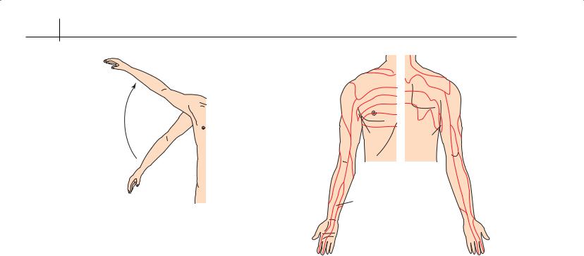
Figure 3.4 Subacromial impingement of the rotator cuff can occur with abduction of the arm. Adapted from Speed et al., 2000
features suggestive of serious underlying pathology in people who present with shoulder pain are shown in Table 3.1.
Rotator cuff disease
Most shoulder complaints (60–70%) are due to rotator cuff disease, a broad term that includes a wide array of diagnostic labels. Some labels derive from clinical features (e.g. painful arc syndrome); some from assumed pathophysiology (e.g. impingement syn- drome—impingement upon the cuff tendons between the acromion and head of the humerus (Figure 3.4) and some from the imaging appearance (e.g. calcific tendinitis, rotator cuff tendinitis or tendinopathy, subacromial bursitis and partialor fullthickness tears).
Based upon MRI scans, asymptomatic cuff tears are common. The incidence increases with age (over half those over 60 years of age have tears), suggesting that it may be part of the normal ageing process combined with repetitive microtrauma. A significant number of asymptomatic tears will become symptomatic over time, and longstanding tears can result in glenohumeral arthritis. Rotator cuff disorders commonly occur in young people engaged in sport involving overhead activities, but are most common in middle and older age. Occupational associations include repetitive movements, working with vibrating tools, working in awkward postures and performing similar work for a prolonged period.
The patient typically complains of pain felt in the shoulder and/or lateral aspect of the upper arm that is worse with overhead activities and at night, particularly when lying on the affected side.
Characteristic features of the examination include pain in the mid-range of active abduction (Figure 3.5) and on resisted shoulder abduction with or without external rotation, and evidence of impingement (production of pain at the anterior shoulder if the arm is flexed forwards to 90 °, adducted and internally rotated, elicited by asking the patient to place their hand on the contralateral shoulder and push up against resistance). In contrast to adhesive capsulitis, which causes global restriction of both active and
|
|
C3 |
C3 |
|
|
|
|
|
|
|
|
C4 |
T2 |
C4 |
|
|
|
||
|
|
T2 |
|
|
|
C5 |
|
|
|
|
T3 |
T3 |
C5 |
|
|
|
T4 |
|
|
|
|
T5 |
|
|
|
|
T2 |
T3 |
T2 |
|
|
T3 |
||
|
C6 |
|
|
|
C5 |
T1 |
|
|
C7 |
|
|
|
||
|
|
|
|
|
|
|
|
|
C5 |
|
|
C7 |
|
|
C6 |
|
|
|
C6 |
|
C8 |
|
|
C8 |
Figure 3.5 Mid or superior painful arcs of abduction represent subacromial impingement or acromioclavicular pathology, respectively
passive movements, passive range of motion is often normal in rotator cuff disease, although certain movements may be restricted by pain. It is therefore important to assess both active (patient moves the shoulder) and passive (the examiner moves the shoulder) movements to distinguish apparent from true restriction of shoulder motion.
Painful weakness and atrophy suggest significant tears. Winging or asymmetry of the scapula may indicate a degree of shoulder instability. The “drop arm” test suggests a complete or large rotator cuff tear.
Calcific tendinitis usually affects women aged 30–50 years and is associated with the formation and resorption of calcific deposits within the cuff. The patient typically presents with acute onset of severe pain, occasionally with a fever and severe limitation of shoulder movements due to pain. In the more chronic stages, pain and catching are reported, and signs of impingement may be noted.
Diagnosis—The diagnosis of rotator cuff disease can usually be made clinically. Blood tests and plain radiographs are not necessary in the absence of “red flags” unless there is a failure to respond to treatment. Plain radiographs may exclude other causes of shoulder pain, such as significant glenohumeral osteoarthritis. If calcific tendinitis is suspected, there may be fluffy calcific deposits, situated just proximal to the rotator cuff insertion (Figure 3.6), and the erythrocyte sedimentation rate and white cell count may also be raised. The diagnostic utility of shoulder ultrasound and MRI in primary care is unknown. Due to the high prevalence of asymptomatic abnormalities in the rotator cuff, these investigations have little to add to the largely conservative management of rotator cuff disease in primary care. Ultrasound and MRI can detect full-thick- ness rotator cuff tears but have less accuracy for detecting partialthickness tears.