Материал: ABC-of-Rheumatology-ABC-Series-
42 ABC of Rheumatology
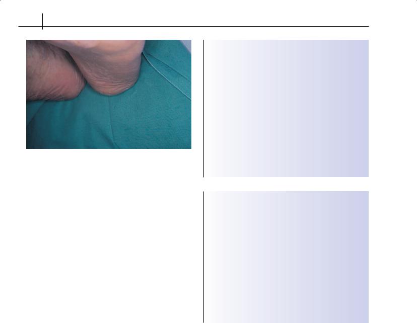
Figure 7.8 Chronic painful plantar heel bursitis
Clinical features—The condition is characterized by an increasingly severe burning, aching and throbbing pain on the plantar surface of the heel. A history of increased activity or weight gain is usual. The heel seems normal but may feel warm. Direct pressure or sideways compression causes pain. The tissues may feel tight and congested.
Treatment—Rest and anti-inflammatory drugs may be useful. Heel cushions and medial arch supports are also used. Stretching exercises (such as rolling a bottle under the foot) can help. Little evidence supports ultrasound treatment, local steroid injections or shortwave diathermy.
Chronic inflammation of the heel pad
This is a distinct clinical condition that usually results from trauma or heavy heel strike. It is sometimes seen in elderly people as their fat pads atrophy or in those who suddenly become more active.
Clinical features—A generalized warm, dull throbbing pain is felt over the weight-bearing area of the heel; this develops over a few months. The pain is most intense typically on first rising. Tenderness is experienced over the heel, which feels tight and distended.
Treatment—Normally, this condition improves with time and rest. Soft heel cushions and medial arch fillers sometimes help. Ultrasound treatment and shortwave diathermy are often used, but controlled trials are few. Steroid injections have an early effect but do not influence the condition’s favourable natural history. Steroid injections can be more painful than the condition unless they are done carefully, with adequate slow infiltration of local anaesthetic (or an ankle tibial nerve block) before injection.
Achilles tendon affections
Inflammation of the Achilles tendon and surrounding soft tissue may be associated with overuse or systemic inflammatory disorders (Box 7.4). Inflammation of the tendon, peritendon tissues and bursae give slightly different clinical pictures. Conditions such as xanthoma can also affect the Achilles tendon and produce fusiform swelling in the tendon. In such cases, cholesterol concentrations
Box 7.4 Achilles tendon affections
Tendinitis
•Presents as painful local swelling of the tendon, which moves with the tendon as the foot is dorsiflexed and plantar flexed
•Important to check the tendon for evidence of partial or complete rupture, which is often missed because of inflammation
•Note recent use of quinolone antibiotics (e.g. ciprofloxacin)
Peritendinitis
•Presents as large diffuse swelling of tissues surrounding the tendon that remains static as the tendon is stretched
•Patients experience pain and crepitus on palpation
Achilles tendon bursitis
•Presents as diffuse fusiform swelling inferior to the Achilles tendon that fills the normal indentation seen below the malleoli and deep to the Achilles tendon
Box 7.5 Common causes of painful heel
Pain within heel
•Disease of calcaneus-osteomyelitis, tumours, Paget’s disease
•Arthritis of subtalar joint complex
Pain behind heel
•Haglund’s deformity (“pump bumps”, “heel bumps”)
•Rupture of Achilles tendon
•Achilles paratendinitis
•Posterior tibial paratendinitis or tenosynovitis
•Peroneal paratendinitis or tenosynovitis
•Posterior calcaneal bursitis
•Calcaneal apophysitis
Pain beneath heel
•Tender heel pad
•Plantar fasciitis
should be checked and treated if raised. Rheumatoid nodules, and occasionally gouty tophi, can also be found within the substance of the Achilles tendon.
Clinical features—Clinical features vary according to the tissues affected. Increased activity leading to an overuse syndrome may be a feature in younger, active patients.
Treatment—Treatment depends on the primary cause. Partial or complete ruptures of the tendon need immobilization and surgical repair. For inflammatory conditions, non-steroidal anti-inflamma- tory drugs may help, as may ultrasound treatment, friction, rest and shock-absorbing heel lifts. Inflammation may be triggered by overuse through poor foot mechanics; in such cases, orthoses may control the pronation. Hydrocortisone injections may be useful if the bursa or peritendons are affected, but they are contraindicated for the tendon itself. Ultrasound imaging may be useful.
A summary of common causes of painful heel is presented in Box 7.5.
Pain in the Foot |
43 |
|
|
|
|
Arthropathies that affect the foot
Osteoarthritis
Osteoarthritis in the foot may be asymptomatic, but it can lead to pain, joint stiffness, functional loss and disability. The most common sites are the first metatarsophalangeal joint (hallux rigidus) and the tarsus joints. Biomechanical factors are often involved in the development of degenerative joint changes (for example, compensatory foot pronation in subtalar osteoarthritis). Trauma, recurrent urate gout, and the demands of fashion—such as inappropriate footwear—are other factors; however, the broad style of modern shoes may be beneficial.
Rheumatoid arthritis
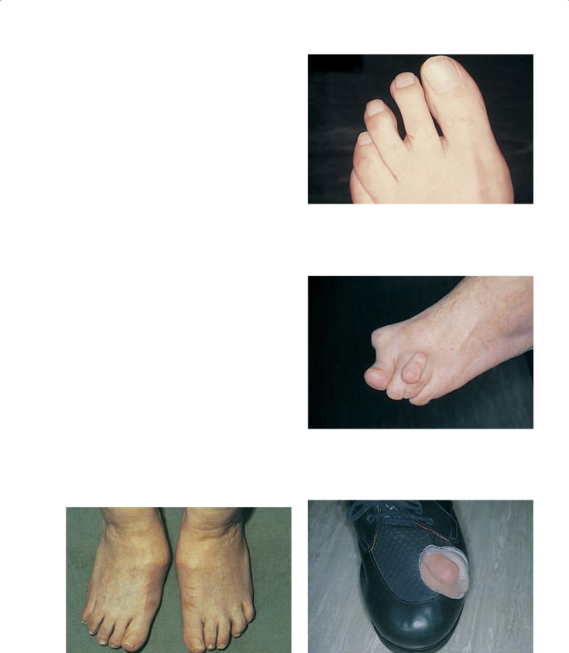
Rheumatoid arthritis often starts in the foot, particularly at the metatarsophalangeal joints. The forefoot is painful and stiff, and direct transverse pressure to the forefoot or squeezing a single metatarsophalangeal joint is painful. Non-specific metatarsalgia is often diagnosed. In the early stages of the disease, the hind foot, particularly the subtalar joint, may also be painful. Synovitis of tendon sheaths around the ankle may also occur. In chronic rheumatoid feet, severe pain in the forefoot may continue, with a sensation of walking on pebbles. Gross deformity causes dysfunction and disability (Figures 7.9–7.12).
Seronegative spondyloarthritis
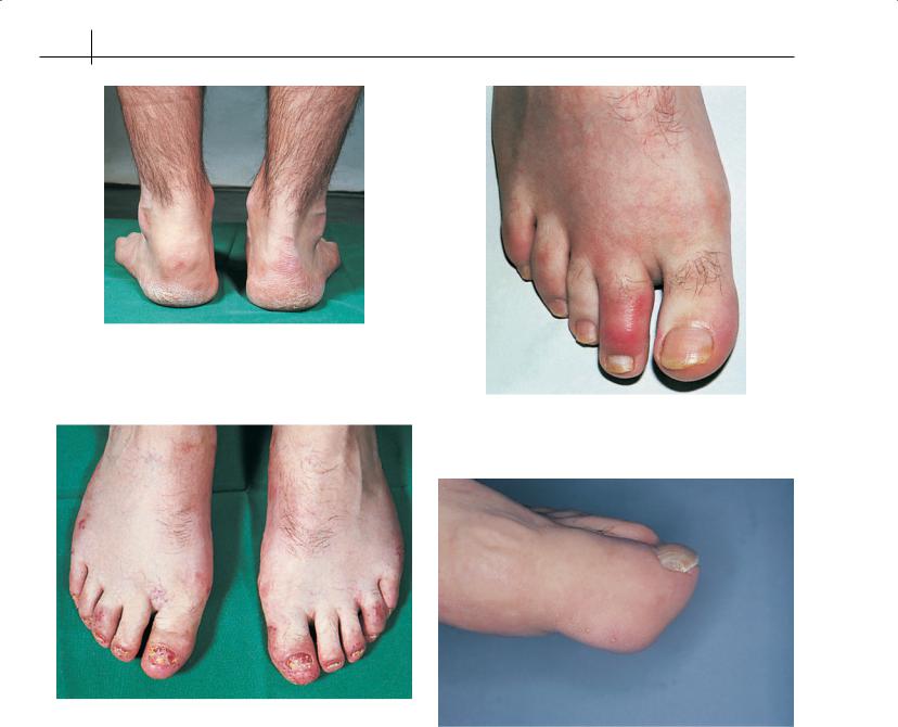
This group includes ankylosing spondylitis (Figure 7.13), psoriatic arthritis (Figure 7.14), undifferentiated seronegative arthropathy and reactive arthritis. Achilles peritendinitis and retrocalcaneal bursitis can be seen. In radiographs, inflammatory spurs may be seen on the calcaneum at the insertion points of the Achilles tendon and plantar fascia. Asymmetrical heel pain may result from a plantar calcaneal enthesopathy.
The pattern of articular involvement in the foot may vary from a single “sausage toe” (dactylitis) (Figure 7.15) to a very destructive arthritis. Painful stiff interphalangeal and metatarsophalangeal joints, often in an asymmetrical pattern, are common. Claw toe
Figure 7.10 Metatarsophalangeal joint synovitis in early rheumatoid arthritis: note a widening of the first and second cleft—the “daylight sign”
Figure 7.11 Extensive foot deformity
Figure 7.9 Midtarsal osteoarthritis |
Figure 7.12 Drastic self-adjustment of surgical shoes to gain pain relief |
44 ABC of Rheumatology
Figure 7.13 Ankylosing spondylitis of the feet: the hind foot is predominantly affected
Figure 7.15 Reiter’s syndrome—“sausage toe” (this is also found with psoriatic arthropathy)
Figure 7.14 Psoriatic arthropathy
Figure 7.16 Large painful tophi over the interphalangeal joint of the hallux
and hallux valgus deformity are more obvious. Nail dystrophy may be seen, with typical psoriatic pitting, onycholysis, subungual hyperkeratosis, discoloration and transverse ridging.
Pustular psoriasis and keratoderma blennorrhagica on the plantar aspect of the foot may contribute to pain when walking.
The diabetic foot—In the presence of neuropathy, the diabetic foot is vulnerable to developing an acute progressive Charcot-like arthropathy. Patients complain of (paradoxically) pain and swelling in the foot, often after minor trauma. The rear and midfoot areas are most often involved. Untreated this will rapidly deteriorate, leaving a disorganized and dysfunctional foot. Treatment must be early and intensive with immobilization of the foot and intravenous bisphosphonates. Early referral is recommended.
Sudeck’s atrophy—A similar condition can develop in the nondiabetic foot following trauma. Sudeck’s atrophy (“reflex sympathetic dystrophy”, or “complex regional pain syndrome type I”) is
a painful condition of the foot and ankle associated with regional bone loss, tissue inflammation and vascular abnormalities. This may be mediated by abnormalities of the autonomic nervous system. The foot may look blue and swollen and is painful at rest and exercise. Plain X-ray may show widespread osteoporosis in the affected area. Treatment is effective pain control and physiotherapy. Sympathetic blockade and intravenous bisphosphonates are sometimes used.
Gout
Chapter 10 discusses the manifestations of acute gout in the foot. In the chronic state, tophi in the foot (Figure 7.16) may ulcerate if they act as pressure points. Permanent destructive joint damage and deformity may result and lead to painful dysfunction in the foot.
Pain in the Foot |
45 |
|
|
|
|
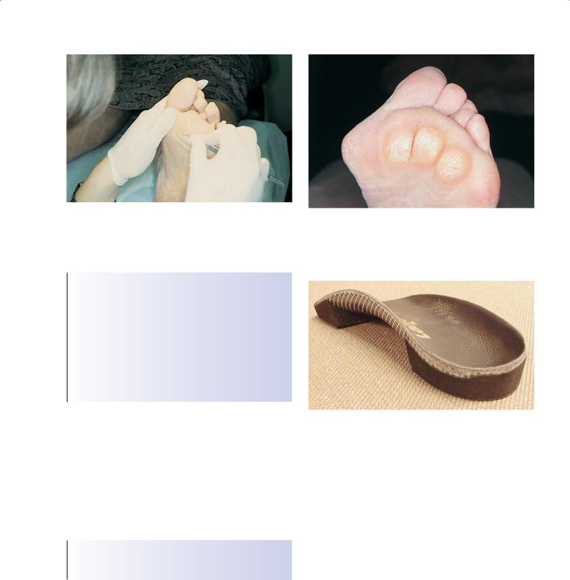
Figure 7.17 Scalpel debridement of plantar lesions provides effective pain relief
Box 7.6 Common abnormalities in the rheumatoid foot
•Hallux valgus
•Lesser toe deformities—e.g. hammer toes and claw toes
•Prominent metatarsal heads with overlying painful callosities or ulceration
•Pronation of foot with valgus heel deformity and collapse of midtarsal joint, giving a flat-footed appearance
•Tenosynovitis, especially of tibialis posterior and peroneal tendons, plantar heel bursitis, calcaneal spur and tendo-Achilles bursitis
•Tarsal tunnel nerve compression syndrome
Figure 7.18 “Walking on pebbles”—metatarsophalangeal callosities in rheumatoid arthritis
Figure 7.19 Custom-made rigid orthoses act as splints to support inflamed joints in early rheumatoid arthritis
Management of rheumatic foot conditions
Patients with rheumatic foot problems (Box 7.6) are best managed by a team that includes a physician, a surgeon and therapists. Podiatrists have a particular role in several aspects of care (Figure 7.17).
Use of orthoses for rheumatic foot problem needs suitable footwear. Podiatrists and orthotists should liaise when extra-depth shoes or surgical shoes are needed
Tissue viability
Joint deformity causes pressure lesions such as callosities (Figure 7.18), corns or ulceration and may be compounded by other factors, such as ingrowing toenails, peripheral neuropathy or the effects of systemic corticosteroids. Podiatrists undertake proce-
dures such as scalpel reduction, design and manufacture of insoles and orthoses, and surgery under local anaesthesia to relieve pain and restore or maintain tissue viability.
Foot function and joint protection
Foot dysfunction due to arthritis can be improved with orthoses, which can be ready-made or individually designed from casts. Orthoses may be used to control deformities—such as the valgus heel seen in rheumatoid arthritis—but they also have a major role in maintaining tissue viability and relieving pain (be it joint, soft tissue or skin lesion in origin) (Figure 7.19). Training towards gait modification may be necessary, and pressure-relieving orthoses of a total contact design may serve to reduce pressures at painful joint sites.
Foot health promotion
Patients will often need advice on daily care of feet. Family members may be involved when patients cannot reach their feet or are unable
46 ABC of Rheumatology
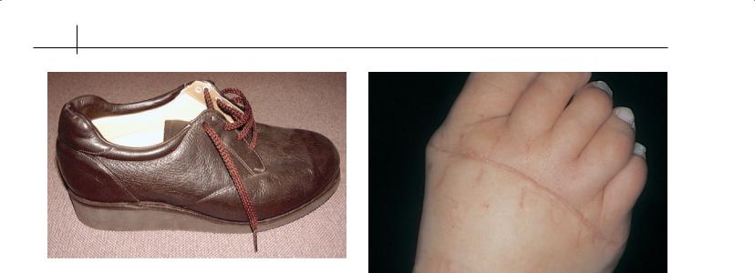
Figure 7.20 Modern stock shoes can be light and comfortable
Figure 7.21 Forefoot arthroplasty is necessary in cases of severe fore foot pain and deformity
to perform tasks on the feet because of other disability. Advice may be needed on splints, walking aids, footwear (Figure 7.20), insoles, foot hygiene and exercise.
Foot surgery
Many rheumatic patients have conditions of the toenails that need surgery under local anaesthetic; they are best dealt with by an experienced clinician such as a podiatrist. Foot surgery may be effective for relieving pain and improving deformity when conservative measures have failed (Figure 7.21).
Further reading
Fam AG. The ankle and foot: regional pain problems. In: Klippel JH, Dieppe PA, eds. Rheumatology. Mosby, London, 1998.
Hintermann B, Nigg BM, Hames MR, Cooper PS, Sammarco GJ, Renstrom PAFH, et al. Foot and ankle. In: Nordin M, Anderson GBJ, Pope MH, eds.
Musculoskeletal Disorders in the Workplace: principles and practice. Mosby, St Louis, 1997: 537–595.
Jayson MIV, Smidt LA, eds. The Foot in Arthritis. Baillière Tindall, London, 1987.
Helliwell PS, Woodburn J, Redmond AC, Turner DE, Davys HJ. The Foot and Ankle in Rheumatoid Arthritis. Churchill Livingstone Elsevier, Oxford, 2007.
Rana NA. Rheumatoid arthritis, other collagen diseases, and psoriasis of the foot. In: Jahss MH, ed. Disorders of the Foot and Ankle: medical and surgical management. WB Saunders, Philadelphia, 1991: 1719–1751.