Материал: ABC-of-Rheumatology-ABC-Series-

Pain in the Knee |
37 |
|
|
|
|
London, 2007. Available online at http://www.clinicalsportsmedicine.com/ chapters/index.htm
Brukner P, Khan K, Crossley K, Cook J, Cowan S, McConnell J. Anterior knee pain. In: Brukner P & Khan K, eds. Clinical Sports Medicine, 3rd edn. McGraw Hill, London, 2007. Available online at http://www.clinicalsportsmedicine.com/chapters/index.htm
Panayi G, Dickson DJ. Clinical Practice Series: Arthritis. Churchill Livingston, London, 2004.
Underwood M. Chronic Knee Pain in the Elderly. Reports on the Rheumatic Diseases Series 5, no. 5. Arthritis Research Campaign, York, UK, 2005. Available online at http://www.arc.org.uk/arthinfo/medpubs/6525/ 6525.asp

CHAPTER 7
Pain in the Foot
James Woodburn1 and Philip S Helliwell2
1Glasgow Caledonian University, Glasgow, UK
2University of Leeds, Leeds, UK
OVERVIEW
•Foot pain is common and can be associated with a number of local or generalized conditions.
•Clinical examination and simple investigations can usually identify the cause of the pain.
•Foot pain is a common feature in most rheumatic diseases, including rheumatoid arthritis and osteoarthritis.
•Podiatrists, general practitioners, rheumatologists and orthopaedic surgeons are involved in the management of foot pain.
Foot pain is common. It may be caused by local disease, be associated with systemic disease or be a reflection of chronic widespread pain. In general, a multidisciplinary approach to treatment is preferable. This is reflected in increasingly close liaison between podiatry, rheumatology and orthopaedic departments. State-registered podiatrists offer a range of treatments, from skin lesion care to orthoses and, more recently, ambulatory forefoot surgery. To understand dysfunction, clinicians should be familiar with the normal development and anatomical variants of the foot (Figures 7.1 and 7.2; Boxes 7.1 and 7.2).
progress to bony ankylosis. The child may be reluctant to push off with the forefoot during walking, and pressure studies show poor contact of the foot to the floor. Lack of use can lead to delayed maturation of bone or soft tissue, and, in such cases, discrepancy in leg length should be sought carefully.
Box 7.1 Characteristics of the adult foot
Three main types of foot
•Normal
•Pronated (flat)
•Supinated (high arch)
Examination
•Examine the foot when bearing weight and when unloaded and be sure to look at the plantar surface of the foot for callus formation (often associated with high pressure)
•Inspect the patient’s shoes for abnormal or uneven wear
•Consult a podiatrist if a structural or mechanical abnormality is suspected—many can be treated with orthoses
Foot pain in children
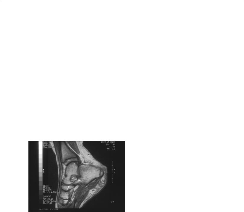
Foot pain may be associated with congenital abnormalities, such as equinovarus deformity. Such structural abnormalities may reflect underlying neurological diseases, such as cerebral palsy. A rigid pronated foot in the early teens may be the first symptom of a tarsal coalition (Figure 7.3). Gait abnormalities, such as intoeing, may be of concern to parents, but they are seldom treated actively.
Juvenile chronic arthritis
The knee and ankle joints are most often affected in all subtypes of juvenile chronic arthritis. Children may present with a limp or reluctance to walk. In the hind foot, pain and reflex muscle spasm can lead to valgus deformity (in two-thirds of cases) or varus deformity (in one-third of cases). In some patients, this may
ABC of Rheumatology, 4th edn. Edited by Ade Adebajo. ©2010 Blackwell Publishing Ltd. 9781405170680.
Box 7.2 Characteristics of children’s feet
Normal foot
•Flexible foot structure (may look flat with a valgus heel)
•Medial longitudinal arch forms when child stands on tiptoe
•Heel-to-toe walking
•Forefoot in line with rear foot
•Mobile joints with painless motion and no swelling
•Adopts adult morphology by about 8 years of age
Abnormal foot
•Inflexible
•Lesser toe deformities
•Rigid valgus (pronated) foot with everted heel position
•High-arch foot with toe retraction and tight extensor tendons
•Toe walking
•Delay or difficulty in walking or running
•Abducted or adducted forefoot relative to heel
•Pain, swelling or stiffness of joints
•Hallux deformity
38
Pain in the Foot |
39 |
|
|
|
|
(a) 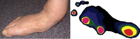 (b)
(b)
Figure 7.1 Abnormally pronated foot (a), with pressure profile showing large weight-bearing surface and higher pressures medially (b)
(a) 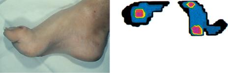 (b)
(b)
Figure 7.2 Abnormally supinated foot (a), with pressure profile showing small weight-bearing surface and high pressures over first and fifth metatarsal heads (b)
Figure 7.3 Tarsal coalition. MRI in a patient with calcaneonavicular coalition. Note synostosis between the calcaneus and navicular bones (arrows)
Pain in the forefoot (metatarsalgia)
Morton’s metatarsalgia (interdigital neuroma)
This normally affects the proximal part of the plantar digital nerve and accompanying plantar digital artery. Trauma to these structures leads to histological changes, including inflammatory oedema, microscopic changes in the neurolemma, fibrosis and, later, degeneration of the nerve. Morton’s neuroma is the result of an entrapment lesion of the interdigital nerve.
Clinical features—Clinical features include a gradual onset, with sudden attacks of neuralgic pain or paraesthesia during walking— often in the third and fourth toe. Examination may show lesser toe deformities, slight splaying of the forefoot, abnormal pronation and hallux valgus. These often occur in women who wear court shoes. Compression of the cleft or laterally across the metatarsal heads may produce acute pain and the characteristic “Mulder’s click.”
Treatment—Patients should be given advice about suitable footwear and possibly should be given orthoses to control abnormal pronation. Injections of local anaesthetic and hydrocortisone around the nerve, or surgical excision, can be helpful.
Stress fracture (march fracture)
Stress fractures are associated with increased activity, and lesions can affect any of the metatarsal shafts, often along the line of the surgical neck. They can occasionally be seen in patients with osteoporosis as a pathological fracture.
Clinical features—Patients have a history of a change in the amount of activity, change in occupation or footwear, or sudden weight gain. The symptom is a dull ache along the affected metatarsal shaft, which changes to a sharp ache just behind the metatarsal head. The pain is exacerbated by exercise and is more acute at “toe off.” Tenderness and swelling is felt over the dorsal surface of the shaft. Pain is produced by compression of the metatarsal head or traction of the toe. X-ray examination may not show the fracture for 2–4 weeks, but if it is important to confirm the diagnosis—e.g. for an athlete who needs advice on whether to continue playing sport—a bone scan can reveal it earlier.
40 ABC of Rheumatology
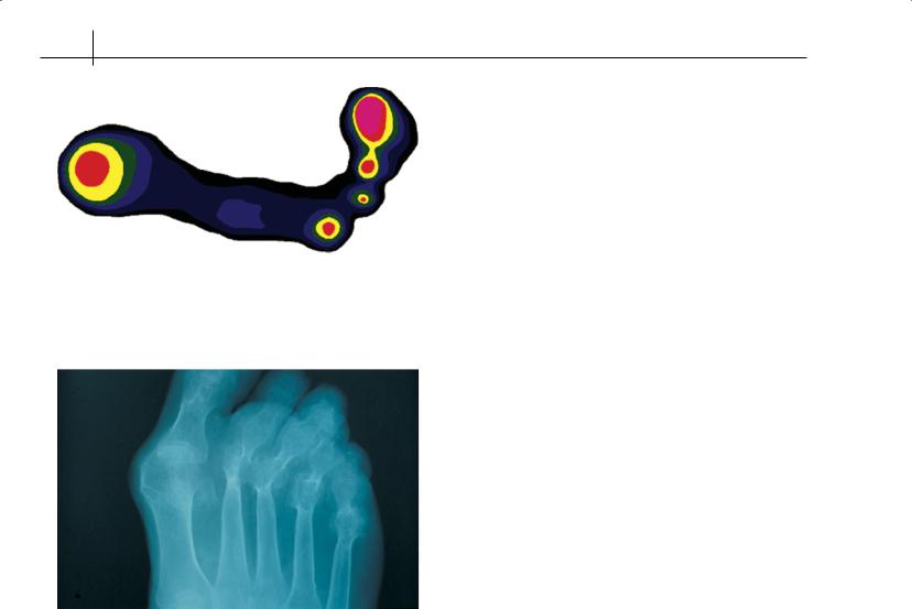
Figure 7.4 Pressure profile of the right foot of a 54-year-old patient with rheumatoid arthritis. Note absent lesser toe contact and high pressure (hot colours) over central metatarsal heads
Figure 7.5 Advanced destruction in the forefoot of a patient with rheumatoid arthritis
Treatment—Rest and local protective padding with partial immobilization are usually enough. These fractures rarely require casting.
Acute synovitis
This condition is normally associated with acute trauma, which leads to inflammation of the synovial membrane and effusion. Freiberg’s disease may also contribute. Systemic causes of acute synovitis, such as rheumatoid arthritis (Figures 7.4 and 7.5) or infection, should be excluded when making a diagnosis.
Clinical features—It is rare in children but often affects young adults. Patients complain of a sudden onset of painful throbbing that is made worse by movement. The patient may have experienced trauma or have a systemic inflammatory disorder. Any movement of the joint produces pain. Fusiform swelling is present around the distended joint, and crepitus may be felt.
Treatment—Rest, immobilization and ultrasound treatment may help if trauma is the cause. Anti-inflammatory drugs sometimes help. Previously unsuspected systemic arthritis should be investigated.
Acute inflammation of anterior metatarsal soft tissue
This common condition is generally found in middle-aged women. It affects the soft tissues of the plantar aspect of the forefoot and is associated with increased shear forces, such as occur when wearing “slip-on” and high-heeled court shoes.
Clinical features—Patients present with a burning or throbbing pain localized to the soft tissues anterior to the metatarsal heads. The pain usually develops over a few weeks, is often associated with walking in a particular pair of shoes, and is usually relieved by rest. The tissues are inflamed, warm and congested. Direct palpation, rotation and simulation of shear forces on the foot exacerbate the pain. Examination of patients’ shoes may reveal a worn insole, with a depression under the metatarsal heads.
Management—Advice on footwear, with adequate support or cushioning, should be given. Associated abnormal pronation or lesser toe deformities should be corrected with orthoses.
Osteochondritis (Freiberg’s infraction)
This quite common condition generally affects the second or third metatarsal heads. It is an aseptic necrosis or epiphyseal infraction associated with trauma and localized minute thrombosis of the epiphysis.
Clinical features—Osteochondritis affects teenagers and is associated with increased sporting activity. The presenting complaint is often a limp, with dull pain associated with movement of the metatarsal phalangeal joint, exacerbated at “toe off ”. The long-term result is a flattened metatarsal head, which can progress to arthritis. The affected joint may be slightly swollen, with a disparity in toe length and width. Traction causes pain. Restricted movement may be due to muscle spasm in the early stages and later to arthritis. Radiographs show distortion of the metatarsal head.
Treatment—In the early stages, rest and immobilization are enough, but sometimes patients eventually need corrective surgery.
Plantar metatarsal bursitis
This condition may affect the deep anatomical or superficial adventitious bursae. In the acute form—such as in dancers, squash players or skiers—the first metatarsal is usually affected, while the second to fourth metatarsals are affected in chronic inflammatory arthritis (Figure 7.6).
Clinical features—Patients present with a throbbing pain under a metatarsal head that usually persists at rest and is exacerbated when the area is first loaded. The acute condition affects men and women equally, usually in younger adults. If a superficial bursa is affected, there will be signs of acute inflammation, with fluctuant swelling and warmth. With deep bursitis, the tissues are tight and congested. Direct pressure or compression produces pain, as does dorsiflexion of the associated digit.
Treatment—Anti-inflammatory drugs are useful; in practice, local gels and systemic oral drugs help. Injections of corticosteroid may
Pain in the Foot |
41 |
|
|
|
|
Figure 7.6 Severe plantar metatarsal bursitis affecting the second metatarsal head of a patient with rheumatoid arthritis. The overlying callus suggests that this is a high-pressure site during normal gait
Box 7.3 Causes of pain in the forefoot
Primary
• Functional and structural forefoot pathologies
Secondary
•Rheumatoid disease
•Stress lesions
•Post-traumatic syndromes
•Diabetes
•Gout
•Paralytic deformity
•Sesamoid pathology
•Osteoarthritis
Unrelated to weight distribution
•Nerve-root pathology
•Tarsal tunnel nerve compression syndrome
Analogous to carpal tunnel syndrome
Often misdiagnosed as foot strain or plantar fasciitis
Primary symptom is burning feeling on sole of foot in the dermatome served by the medial plantar nerve
be indicated in severe cases. Patients must rest the affected part; this may be achieved by protective padding. Any underlying deformity or foot type with abnormal function should be assessed and treated.
A summary of causes of pain in the forefoot is presented in Box 7.3.
Plantar fascia affections
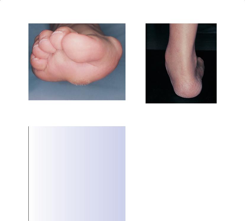
Pain along the medial longitudinal arch is quite common. Most affected patients have abnormal foot mechanics, such as abnormal pronation, valgus heel (Figure 7.7) or flat foot. Mechanical dys-
Figure 7.7 Valgus heel with bulging of the talar head medially
function and change in medial arch posture can place strain on soft tissues, which results in localized or more diffuse pain—the foot’s equivalent to low back pain syndrome. Other conditions include true plantar fasciitis, which is characterized by a few fast-growing nodules in the fascia, and plantar fibromatosis, which is characterized by fibrous nodules and contracture of the fascia.
Treatment of true plantar fascial strain requires rest, control of abnormal function with orthoses, and stretching exercises. Ultrasound treatment seems helpful, but controlled trials are lacking.
Painful heel
Sever’s disease (calcaneal apophysitis)
This was thought to be an avascular necrosis of growing bone but is now interpreted as a chronic strain at the attachment of the posterior apophysis of the calcaneus to the main body of the bone, possibly from pull of the Achilles tendon. It is analogous, therefore, to Osgood-Schlatter disease of the tibial tuberosity.
Clinical features—The condition usually affects boys aged 8–13 years, who complain of a dull ache behind the heel of gradual onset that is exacerbated by jumping or occurs just before “heel lift”. A limp is usually seen with early heel lift. Rest normally relieves the pain. Tenderness is seen over the lower posterior part of the tuberosity of the calcaneus. Radiographs are usually normal.
Treatment—In most cases, reassurance and advice about reducing activities will suffice: the condition usually subsides spontaneously. In some cases, heel lifts help; occasionally, if the pain is severe, a below knee walking cast is needed.
Plantar calcaneal bursitis (“policeman’s heel”)
This is inflammation of the adventitious bursa beneath the plantar aspect of the calcaneal tuberosities (Figure 7.8). It is associated with shearing stress caused by an altered angle of heel strike.