Материал: ABC-of-Rheumatology-ABC-Series-

CHAPTER 22
Sports and Exercise Medicine
Cathy Speed
Cambridge University Hospital, Cambridge, UK
OVERVIEW
•Sports and exercise medicine addresses the prevention and management of activity-related medical complaints, and the use of exercise for health-related benefit.
•Exercise and increased activity have proven benefits, both in the prevention and treatment of a wide range of conditions.
•Assessment of any sports injury requires an understanding of the potential intrinsic and extrinsic aetiological factors.
•Absolute rest is rarely if ever a component of a structured rehabilitation programme, which should aim to maintain physical fitness while restoring normal function.
•Increased activity can be beneficial to all, and be achieved by all, and it is incumbent on all health-care professionals to promote increases in physical activity.
Introduction
Sports and Exercise Medicine addresses the prevention and management of sportsand activity-related medical complaints, and the use of exercise for health-related benefit. Rheumatologists are often faced with sports injuries and have many patients who will benefit from an exercise prescription.
Sports injuries
Introduction
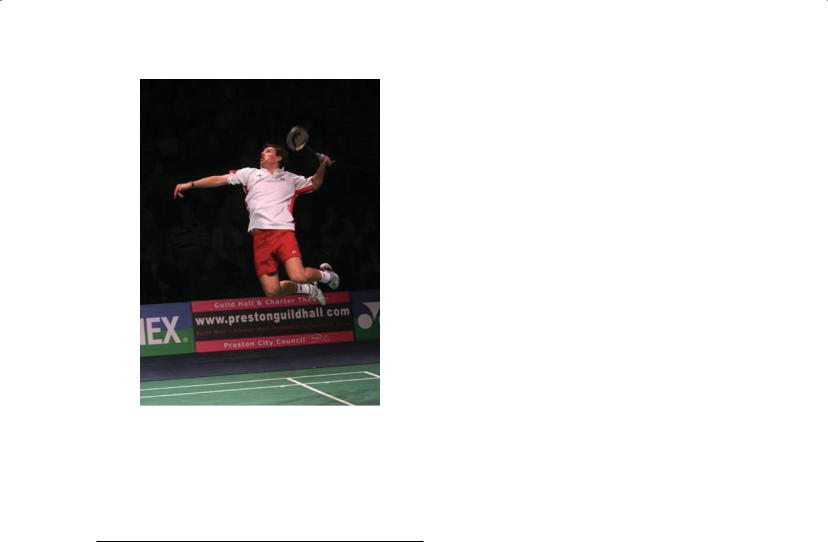
The key to managing sports-related injury is having an understanding of the patient and their sport. As with any patient, it is important to consider the patient’s ideas, expectations and concerns. Those with an “athletic psyche” may have high anxiety levels about their injury and its implications, unrealistic expectations for recovery goals and time frames and a tendency to “overcomply” with rehabilitation programmes. An insight into the mechanics, training and techniques of the sport involved is also important, as this allows the underlying cause of the injury to be addressed (Figure 22.1).
ABC of Rheumatology, 4th edn. Edited by Ade Adebajo. ©2010 Blackwell Publishing Ltd. 9781405170680.
Assessment
When assessing sports injuries, it is helpful to consider intrinsic and extrinsic factors (Table 22.1).
Intrinsic factors encompass physical, physiological and psychological aspects of an individual that may contribute to injury. Importantly, what may be considered “abnormal”—for example, asymmetry of muscle development or joint range of motion—may be normal in relation to a trained athlete. Similarly, what is normal in the general population may be abnormal for an athlete—for example, average flexibility in a gymnast is likely to be abnormal.
Extrinsic factors play a significant role in the development of injury. Doing “too much, too soon, too often” is a common error in athletes of all levels. Other factors, such as inappropriate or recent change in equipment, environmental conditions and competing surfaces, also may play a role.
A central concept in the assessment of an athlete, in particular when considering injury, relates to the delicate balance that exists between optimal mobility of a joint or a series of joints, and optimal stability. Frequently this balance is disrupted in the development of injury and must be considered in diagnosis and treatment of any athletic complaint.
History—The history addresses the injury, training and competing habits, the potential role of other extrinsic factors, previous injury history and other medical issues. The mechanism of injury is important in elucidating the diagnosis, as it will implicate the structures involved and the severity of the injury.
Pain is most frequently the cardinal symptom and a usual pain history is taken: its site(s), radiation, timing of onset and subsequent temporal pattern, aggravating and relieving features and associated symptoms. The degree of swelling and its rapidity of onset after injury frequently correlate with the severity of injury. Instability or a feeling of “pre-instability” are highly relevant in sport and may indicate a true structural deficit or a lack of neuromuscular control. Clicking and clunking of a joint is relevant, particularly if new or painful. Neurological symptoms may be present and may indicate a true neurological deficit or, more frequently, neural irritation in association with a chronic soft-tissue injury.
A history of treatments used to date, a medication history (including vitamins and supplements) and a general medical background are all important. For example, underlying medical com-
142
Sports and Exercise Medicine |
143 |
|
|
|
|
Figure 22.1 An insight into the mechanics, training and techniques of the sport is important in understanding sports injuries. This figure demonstrates the demands of a sport such as badminton, and the fine balance that exists between mobility and stability is a central concept in the consideration of sports injuries. Figure courtesy of Badminton England
Table 22.1 Common extrinsic and intrinsic factors in sports injuries
Intrinsic |
Extrinsic |
|
|
Hypermobility |
Training: too much, too |
|
soon, too often |
Muscle weakness/imbalance |
Technique |
Poor flexibility (local, general) |
Equipment |
Femoral anteversion |
Surface |
Tibia varum/valgum |
Environment |
Pes planus/cavus |
Drugs (e.g. anabolic steroids/ |
|
corticosteroids) |
Presence of another injury |
Poor nutrition |
Chronic diseases (e.g. rheumatoid arthritis) |
|
|
|
plaints such as seronegative arthropathies will not infrequently masquerade as sports injuries, and other serious disorders including tumours should always be considered in an individual with regional musculoskeletal pain. In all athletes, underlying issues relating to bone health should always be considered. The age of the patient is also very important: children have fragile skeletons with vulnerable growth plates and an increased risk of avulsion injuries. The senior population have an increased susceptibility to softtissue injuries and the influence of co-morbidity requires consideration in diagnosis and management.
Examination—Examination commences with a general examination, in particular looking for stigmata of other disease, hypermo-
bility and assessment of the spine, as dysfunction here can contribute to injury. Assessment of asymmetry of muscle groups, flexibility and joint range of motion, is important but must be interpreted carefully. Core stability and control—the ability to control the body adequately during movement—should be assessed, as it is so often lacking in the injured athlete. Regional assessment of the injury follows the usual strategy of “look, feel, move and special tests”. Identification of the site(s) of tenderness, swelling, instability and neurovascular status follows.
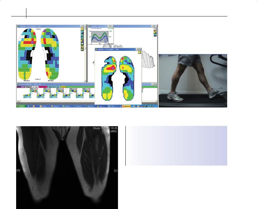
Although assessment commences with examination of the patient at relative rest, it is very important to proceed to dynamic assessment where there is any doubt about the nature and cause of the injury (Figure 22.2). It may be necessary to evaluate the individual during or after a rigorous set of exercises in order to reproduce symptoms. Video analysis may be very informative in illustrating the underlying factors contributing to injury; input from a coach or technical expert is also often helpful.
Investigations—Investigations (imaging in particular) are frequently required in the assessment of the injury, but should be requested only after a clinical diagnosis is made and interpreted carefully. No imaging is foolproof, and it is vital to request the correct test for the suspected injury.
Imaging includes plain X-rays, diagnostic ultrasound, magnetic resonance imaging (MRI), computed tomography (CT) and isotope bone scans. Plain X-rays assess for fractures, myositis ossificans, loose bodies and underlying joint damage but are not sensitive to early stress injuries. Stress views may be necessary to assess for instability. Diagnostic ultrasound demonstrates soft-tissue anatomy and impingements and allows dynamic assessment of the joint in question. MRI provides further information of the surrounding anatomy, bone oedema and some soft-tissue injuries (Figure 22.3), but MR arthrography is necessary to evaluate the labra of shoulder and hip most accurately. CT scanning for loose bodies, and scintigraphy for stress injuries in particular, may be indicated. Laboratory investigations for underlying medical complaints may be necessary.
Compartment studies, involving measurement of muscle compartment pressures before, during and after exercise are important in the evaluation of individuals with possible chronic exertional compartment syndromes.
Other investigations, such as dual X-ray absorptiometry scanning for those with recurrent stress fractures may be warranted. The sites of low bone density in athletes may differ from the general population in view of the different patterns of skeletal loading; scanning of sites such as the forearm is often necessary.
Management
The management of sports related injuries commences with an accurate diagnosis and identification of all the contributing factors. Education and counselling in relation to the injury, and discussion and agreement on an appropriate management strategy are vital. Appropriate levels of compliance will be enhanced by ensuring the athlete has a clear understanding of the injury, its implications and treatment. Clear goals need to be set, and reviewed regularly. Pain
144 ABC of Rheumatology
Figure 22.2 Gait analysis and shoe pressure measurement can be particularly helpful in the assessment of lower limb injuries
Box 22.1 Principles of management of sports injuries
• Early diagnosis, identify and correct the mechanism
• In the acute phase: PRICES
• Control pain in order to allow rehabilitation to proceed
• Rehabilitation addresses flexibility, strengthening, proprioception, sports-specific work such as agility, speed, power, technique
• Graduated return to sport
Figure 22.3 MRI of thighs showing left hamstrings muscle injury
management is important, principally to allow rehabilitation to proceed. In the acute injury, the classical PRICES regime (protect, rest, ice, compression (if necessary), elevation, support) is followed. Rest is relative; the unaffected areas can and should continue to be exercised; for example, swimming or aquajogging after a tibial stress fracture. Supports and braces—such as a splint in ankle sprain, or boot in stress fractures of the foot—enable the individual to mobilize without overstressing the site of injury.
The most important aspect of management is rehabilitation, which, after the PRICE regime if necessary, addresses joint range of motion, proprioception, flexibility and strength issues initially. Underlying asymmetries in strength and flexibility are focused on, core stability is addressed and the patient then moves towards sports specific rehabilitation to include power, agility and control during appropriate activities (Box 22.1).
Pain control may be necessary in order to allow rehabilitation to proceed. This may be in the form of ice/heat modalities, simple analgesics or non-steroidal anti-inflammatory drugs. Injections may be useful. For example, local anaesthetic may be used to identify the source of pain, and corticosteroid for chronic injuries in which inflammation is ongoing. Injudicious loading under the influence of analgesia, and in particular corticosteroid, must be avoided.
Surgery may be required—either early, or if other management approaches fail. The decision to intervene operatively will depend upon the nature of the injury and the circumstances of the athlete. For example, elite athletes may choose to have surgical intervention in the hope it will speed recovery to promote a swift return to sport. Surgery is never an isolated treatment; rehabilitation remains an essential part of management. Examples of indications for early surgical intervention include fractures, acute traumatic tendon ruptures, significant loose bodies and labral injuries, and exertional compartment syndromes.
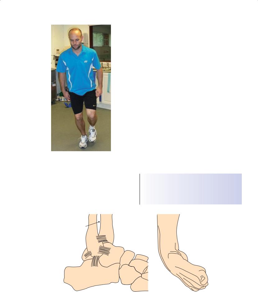
Even when surgery is likely to be indicated, many injuries may be managed in the initial phases with rehabilitation (Figure 22.4). This may be termed “pre-habilitation”: where strength and proprioception can be partly restored, enhancing the pace of postoperative recovery.
Sports and Exercise Medicine |
145 |
|
|
|
|
Figure 22.4 Rehabilitation involves progression from basic flexibility and strength exercises to sports-specific activities. Here, the athlete performs a single leg squat, a simple core stability exercise. Figure courtesy of Badminton England
Common sports injuries—the acute injury
Sports injuries can be broadly divided into acute injury and chronic overuse injury. The most common acute injuries involve ligaments (sprain) or muscles (strain) and vary enormously in severity in terms of the extent of injury (from a simple sprain/strain to a
complete rupture), the muscle or ligament affected (e.g. a straightforward long head of biceps tear to the problematic hamstring) and the location within the muscle/tendon complex (a midsubstance tear compared with a tear at the musculotendinous junction). The impact of any injury is of course further complicated by the functional aspirations of the individual and their age, which will affect the site of injury and the potential for healing. The most common acute sports injury is undoubtedly the ankle sprain.
Ankle sprain—Inversion injuries to the lateral ligament complex of the ankle are one of the most common causes of long-term disability after injury. Injuries initially occur in plantar flexion and slight inversion, such as at push off (Figure 22.5). Recurrent injuries can occur with minimal trauma, e.g. slipping on a kerb, indicating instability: functional (muscle weakness, loss of proprioception) or mechanical (significant ligament disruption).
After the acute injury, the degree of soft-tissue damage can be estimated by the extent of the swelling, and the likelihood of bone injury by clinical features (Box 22.2). Ankle sprain can result in additional damage that may cause either ongoing instability or chronic pain (Box 22.3). Popping, clicking, locking and neuralgia may all be significant.
Clinical assessment includes assessment of balance and proprioception, mechanical stability, sites and degree of tenderness, and neurovascular status. Management of the acute injury should focus on early mobilization, range of motion and strengthening exercises (particularly the peroneals) and proprioceptive work. Use of an ankle brace may help in an earlier return to sport.
Box 22.2 Ottawa Ankle Rules: when to X-ray for bony injury after ankle sprain
•Inability to bear weight and/or
•Bone tenderness at the posterior edge of the tibia or fibula or tip of either malleolus
Fibula |
Tibia |
Intraosseus
membrane
ATFL (tibiofibular)
ATFL (talofibular)
PTFL
Talus
CFL
Calcaneus
(a) |
(b) |
Figure 22.5 An ankle sprain involves a tear to one or more of the lateral ligaments of the ankle (a) and usually occurs with the foot in plantar flexion in slight inversion (b).
146 ABC of Rheumatology
Box 22.3 Causes of pain and instability after ankle sprain
Articular injury
•Chondral/osteochondral fracture
•Meniscoid lesion
Bony injury
• Fibula
Nerve injury
• Superficial peroneal/posterior tibial/sural
Tendon injury
•Tibialis posterior (tear, tendinosis)
•Peroneal (subluxation/dislocation/tear/tendinosis)
Ligament injury
•Mechanical instability due to lateral ligament damage
•Syndesmosis/subtalar joint
Impingement
• Anterior osteophyte/anteroinferior tibiofibular ligament
Miscellaneous
•Failure to regain normal motion (tight Achilles tendon)
•Proprioceptive deficit with repetitive sprains (functional instability)
Box 22.4 Components of an exercise prescription
•Aerobic
Activities selection
Duration
Frequency
Intensity
•Resistance training
•Flexibility training
Address issues such as adverse biomechanics from osteoarthritis before commencing; counselling, supervision and progression of programme
Common sports injuries—the chronic/overuse injury
Whereas acute injuries are more common while working in a competitive sporting environment, the injury that presents most commonly to a sports medicine clinic is the overuse injury. Overuse injuries are defined by the inability of a normal structure to cope with an excessive load, as opposed to an insufficiency injury, in which a pathologically weak structure is unable to cope with a normal load. Overuse injury can affect the tendon (e.g. tendinopathy of which Achilles, patella and common elbow extensor and flexor are common), muscle (examples of which are chronic exertional compartment syndrome and medial tibial stress syndrome) and bone. The classical overuse bone injury is the stress fracture, the most common sites being the tibia, metatarsals and lumbar spine (spondylolysis), but they can occur in any loaded bone (e.g. ribs in rowers).
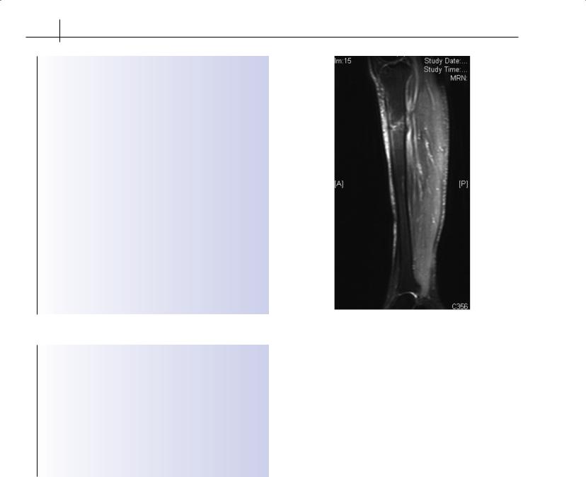
Figure 22.6 MRI showing a severe stress fracture of the proximal tibia
Stress fractures—Stress injuries to bone are a common reaction to repetitive loading of the skeleton without adequate time for remodelling. Although most stress injuries are fatigue-related, the possibility of insufficiency fractures, particularly in lightweight athletes, must always be considered. Most stress fractures will respond to relative rest, support and correction of the underlying cause (training, biomechanics, equipment errors). However, certain stress fractures are associated with an increased risk of poor healing/ completion, including the superior surface of the femoral neck, anterior tibial cortex and navicular. These are areas that are under tension (rather than compression) and/or have poor vascular supply. They are managed either by non-weight-bearing and close monitoring or early surgical intervention (Figure 22.6).
Exercise prescription
The benefits of exercise in the prevention and management of disease are well established. Many patients with rheumatological diseases should be given an exercise prescription, as many are at increased risk of medical complications such as osteoporosis and cardiovascular events. Current recommendations are that adults aged 18 to 65 years need moderate-intensity aerobic physical activity for a minimum of 30 minutes on 5 days each week or vigorousintensity aerobic physical activity for a minimum of 20 minutes on 3 days each week and strengthening exercise two to three times weekly. This may need to be modified for those with diseases such as rheumatoid arthritis, but provides a target.
The exercise prescription has a number of components (Box 22.4), which are adjusted according to the individual’s needs, char-