Материал: ABC-of-Rheumatology-ABC-Series-
|
|
|
|
Is it a Connective Tissue Disease? |
137 |
|
|
|
|
|
|
|
|
|
|
|
|
|
|
|
|
|
|
|
|
|
|
|
|
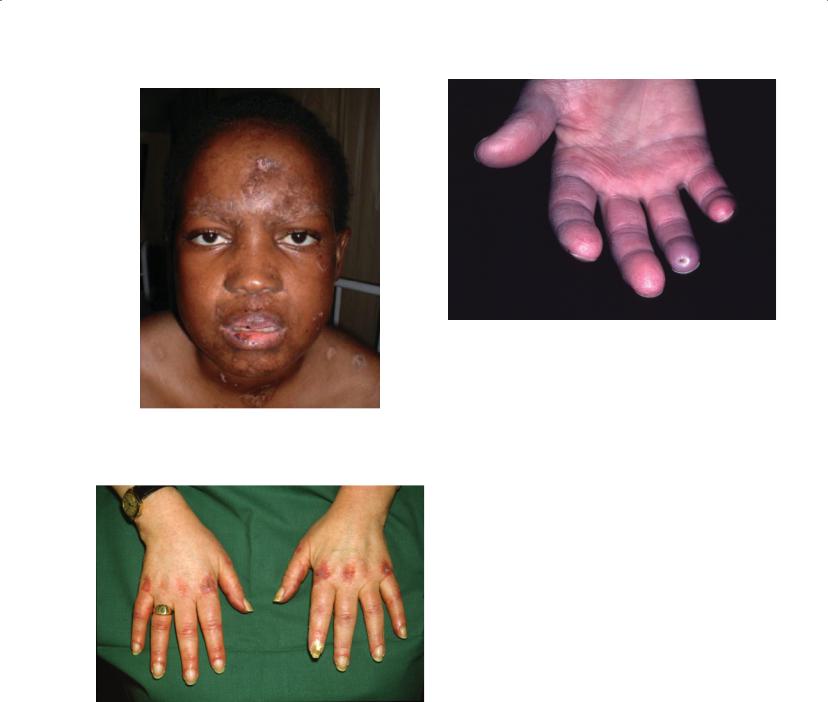
Figure 21.2 Active discoid lupus erythematosus
Figure 21.3 Hands in dermatomyositis
characteristic patterns associated with the different diseases. Early diagnosis is aided by recognition of distinctive serological profiles that are generally present with the earliest clinical manifestations. Diagnosis can also be facilitated by typical laboratory abnormalities and histological changes in the tissues involved. For example, microscopic polyangiitis, which presents with weight loss, fever, polyarthritis and active urinary sediment, can be distinguished from lupus by an autoimmune response characterized by pANCA antibodies directed against myeloperoxidase and the typical histological picture of pauci-immune focal necrotizing glomerulonephritis. Similarly, dermatomyositis sine myositis, which presents with photosensitive eruptions on the face, arms, and hands and is
Figure 21.4 Finger pulp scars from previous digital ischaemic ulceration in a patient with systemic sclerosis
associated with myalgia, can be distinguished from lupus by the distribution of the eruption, a raised serum creatine kinase, and typical changes on muscle biopsy, despite the absence of frank weakness (Figure 21.3).
Diagnosis is often complicated if lupus is part of an overlap syndrome and the patient fulfils classification criteria of more than one connective tissue disease (as opposed to undifferentiated connective tissue disease). The most common overlaps with systemic lupus erythematosus are patients who also have features of systemic sclerosis (Figure 21.4), polymyositis, or both, and patients with rheumatoid arthritis. Sometimes patients present with an overlap syndrome; at other times, the picture evolves sequentially. Development of Sjögren’s syndrome during the course of systemic lupus erythematosus is well established, but occasionally patients with primary Sjögren’s syndrome develop typical features of lupus, especially photosensitive eruptions typical of subacute cutaneous lupus erythematosus, after many years of disease.
Patients with an overlap of systemic lupus erythematosus and scleroderma or polymyositis, or both, often have a distinctive serological profile that includes high levels of antibodies to U1RNP. These patients have been suggested to have a distinctive connective tissue disease “mixed connective tissue disease.” This concept is very controversial, however, with critics and protagonists.
A number of series report patients who fulfil criteria for both systemic lupus erythematosus and rheumatoid arthritis. For these patients, the term “rhupus” has been coined. These patients are usually easier to recognize when the rheumatoid arthritis develops first, but they are characterized ultimately by typical rheumatoid features such as erosive arthritis, subcutaneous nodules and rheumatoid factor. These features are accompanied by cutaneous, renal, haematological and other clinical manifestations characteristic of systemic lupus erythematosus, but unusual for rheumatoid arthritis, with the presence of anti-nDNA antibodies.

138 ABC of Rheumatology
Differential diagnosis of connective tissue diseases
A common clinical conundrum is the distinction of systemic lupus erythematosus from other connective tissue diseases (Table 21.4). All frequently present with a mixture of systemic symptoms, including fever and weight loss, and musculoskeletal and/or mucocutaneous involvement. The combination of a careful history and physical examination, urine analysis, chest X-ray, laboratory tests for an acute-phase response, blood count, serum biochemistry, complement levels, creatine kinase and serological profile, however, results in the correct diagnosis in a high proportion of cases.
Drug-induced lupus
A carefully elicited drug history is essential to exclude drug-induced lupus. The management of this is very straightforward, involving discontinuation of the offending agent and short-term anti-inflam- matory treatment. Procainamide and hydralazine carry the highest
risk of inducing a lupus-like syndrome but are now seldom prescribed in clinical practice. In more recent years, several cases of drug-induced lupus have been reported in association with minocycline, a drug is prescribed often for acne, and sulfasalazine, a disease-modifying antirheumatic drug in rheumatoid arthritis, although individual risk of drug-induced lupus is low with these agents. In the context of rheumatoid arthritis, the diagnosis is sometimes difficult to make, particularly as antinuclear antibodies are present in up to 50% of patients with the condition. Druginduced lupus is rare in people of African origin. The clinical presentationissimilartothatof idiopathicsystemiclupuserythematosus, with systemic features including fever and weight loss, arthralgia or frank arthritis, and serositis (particularly common with procainamide). Major organ involvement, such as nephritis and central nervous system manifestations, is less common, although renal disease can rarely occur in sulfasalazine-induced lupus. A high level of antinuclear antibodies usually shows a homogeneous pattern from the earliest presentation, and the typical preponder-
Table 21.4 Distinguishing features of connective tissue diseases
Condition |
Distinguishing features |
|
|
Systemic rheumatic diseases
Rheumatoid arthritis
Systemic sclerosis Dermatomyositis Primary vasculitis Behçet’s syndrome Adult Still’s disease
Hereditary periodic fever syndromes (familial Mediterranean fever, TNF-associated periodic syndrome, hyperimmunoglobulin D, etc)
Prominent signs of synovitis; multisystem involvement uncommon at presentation; no autoantibodies associated with connective tissue disease
Pronounced Raynaud’s phenomenon; scleroderma; characteristic serological profile Distinctive pattern of eruption; prominent muscle involvement; serological profile Distinctive renal involvement; neutrophilia (sometimes eosinophilia); serological profile Lack of typical serological features
Typical fever pattern; lack of typical serological features
Intermittent manifestations; no autoantibodies associated with connective tissue disease
Other systemic disorders |
|
Autoimmune hepatitis |
Typical liver involvement; absence of typical lupus features |
Sarcoidosis |
Typical histology; no autoantibodies associated with connective tissue disease |
Histiocytic necrotizing lymphadenitis (Kikuchi–Fujimoto’s |
Typical histology; lymphadenopathy, fever, neutropaenia and occasional antinuclear |
disease) |
antibodies |
Angioimmunoblastic lymphadenopathy |
Typical histology; no autoantibodies associated with connective tissue disease |
Other causes of photosensitivity and red face |
|
Polymorphous light eruption |
Lack of systemic features; different histology; absent autoantibodies |
Rosacea |
Papulopustular eruption; non-systemic; absent autoantibodies |
Seborrhoeic dermatitis |
Different morphology and histology; non-systemic, absent autoantibodies |
Contact dermatitis |
History of allergen contact; pseudovesicle; no autoantibodies |
Jessner’s benign lymphocytic infiltration |
Typical histology; negative serology |
Erythrohepatic protoporphyria |
Vesicobullous lesions; urinary and plasma porphyrin profile; no antibodies associated with |
|
systemic lupus erythematosus |
Syphilis |
Typical histology; diagnostic serology |
Lupus vulgaris |
Painful nodular cutaneous form of tuberculosis |
Other causes of fatigue and musculoskeletal pain |
|
Fibromyalgia |
No objective inflammation; no autoantibodies associated with connective tissue disease |
Hypothyroidism |
Little objective inflammation. May have Raynaud’s phenomenon and carpal tunnel syndrome |
|
|
Is it a Connective Tissue Disease? |
139 |
|
|
|
|
ance of anti-histone antibodies can be shown with specific assays. Antibodies to native DNA and “extractable nuclear antigens” commonly associated with idiopathic systemic lupus erythematosus are invariably negative, except in the case of minocycline-induced lupus. The gold standard for diagnosis of drug-induced lupus, however, is that it resolves after the drug is stopped; the symptoms improve within days to weeks, although the antinuclear antibodies may take a year or two to disappear.
The anti-TNF agents, mainly used in the treatment of rheumatoid arthritis, but also for Crohn’s disease and ankylosing spondylitis, also induce antinuclear antibody and, specifically, anti-nDNA antibody production. This phenomenon has been observed with all of the anti-TNF agents currently on the market, although more often with infliximab than adalimumab or etanercept. Only a very small proportion of patients develop a lupus-like illness, manifesting mainly with skin rashes, worsening polyarthritis and serositis. In all cases the illness has been reported to be mild and has resolved on discontinuation of the anti-TNF agent. As in the case of sul- fasalazine-induced lupus, diagnosis can be challenging in patients with rheumatoid arthritis who have pre-existing antinuclear antibodies.
Other disorders of the skin
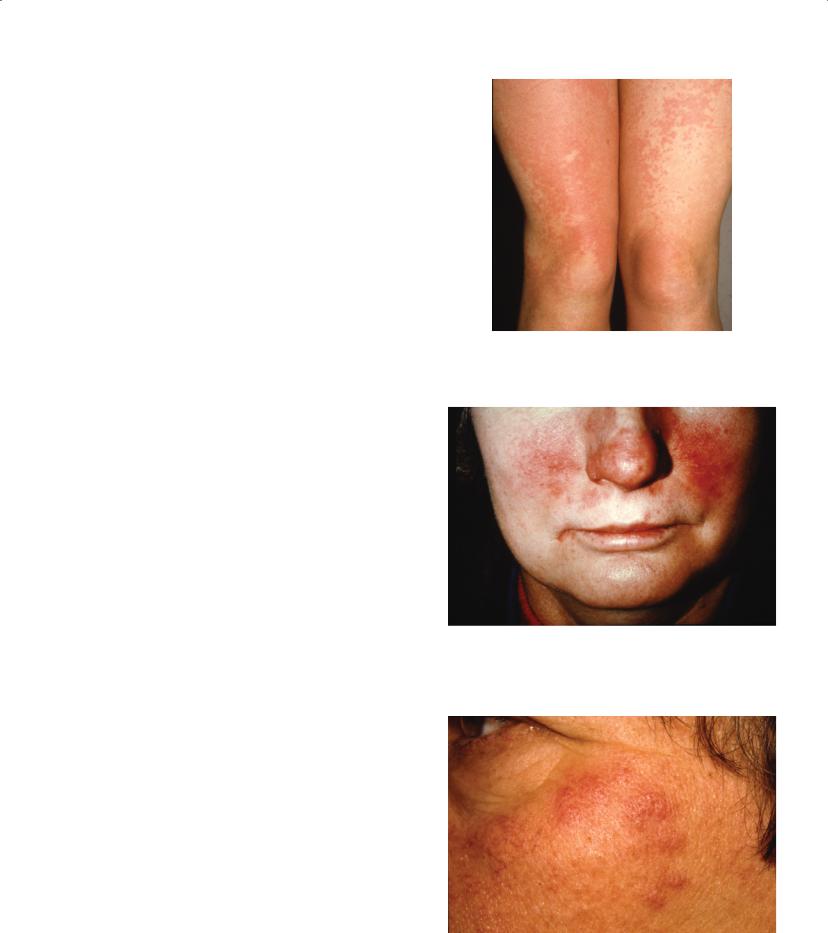
One of the most common conundrums in the connective tissue disease clinic is the patient referred with a history of photosensitivity or red face in association with musculoskeletal symptoms and, perhaps, systemic features such as fatigue. Photosensitive eruptions are common in the normal female population or may be induced by, for example, non-steroidal anti-inflammatory drugs. About 10% of women develop polymorphous light eruption—a pruritic papular eruption that occurs within hours of sun exposure, typically on normally covered sites, that spares the face and hands, and that resolves within days without epidermal change (Figure 21.5). In contrast, photosensitivity in systemic lupus erythematosus also affects the face and hands. The latent period after sun exposure is usually longer, the skin is less pruritic, and the eruption persists longer.
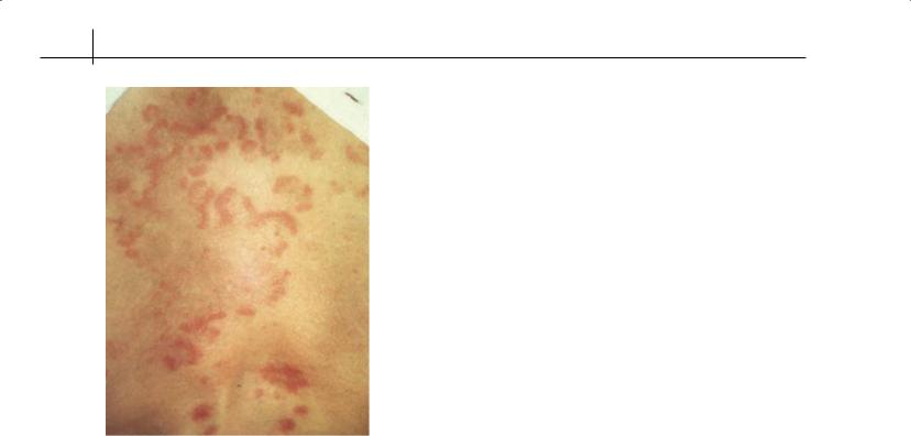
Similarly, the facial erythematous rash that is seen typically in patients with systemic lupus erythematosus must be distinguished from other causes. Typical rosacea consists of papulopustular lesions on a background of telangiectasia (Figure 21.6). Sometimes, light exposure aggravates this condition and a biopsy is sometimes needed to distinguish atypical forms from lupus. Benign lymphocytic infiltration, such as Jessner’s (Figure 21.7), may produce papular or annular lesions that are indistinguishable clinically from subacute cutaneous lupus erythematosus (Figure 21.8) and tumid (papular) lupus erythematosus. The typical histological appearance includes a dense dermal lymphocytic infiltrate without the characteristic epidermal changes of lupus. Seborrhoeic dermatitis may affect the cheeks and paranasal folds and is usually pruritic and associated with desquamation. Contact dermatitis, which may be caused by cosmetics, produces superficial erythema, pseudovesicles and sometimes eyelid swelling. Lupus vulgaris, a painful nodular cutaneous form of tuberculosis, often affects skin over the nose and ears.
Figure 21.5 Polymorphous light eruption
Figure 21.6 Rosacea papules and pustules
Figure 21.7 Papular light eruption and Jessner’s
140 ABC of Rheumatology
Figure 21.8 Rash in subacute cutaneous lupus erythematosus
Fibromyalgia
Fibromyalgia syndrome (see Chapter 8) is often mistaken for lupus, especially if the test for antinuclear antibodies is also positive, and sometimes is treated inappropriately (e.g. with corticosteroids). In addition, a significant proportion of people with fibromyalgia have other features that could be interpreted as manifestations of a connective tissue disease such as Raynaud’s phenomenon, sicca symptoms and cognitive dysfunction. In those mistakenly treated with corticosteroids, although no evidence shows therapeutic efficacy in fibromyalgia, steroid withdrawal can make the symptoms worse. No evidence exists of an increased prevalence of positive antinuclear antibodies or the occurrence of connective tissue disease in patients with fibromyalgia. It has become increasingly apparent, however, that patients with connective tissue disease—especially those with systemic lupus erythematosus and Sjögren’s syndrome—have a high prevalence of fibromyalgia that makes a considerable contribution to morbidity but is unrelated to the activity of the disease.
Endocrine disorders
Hypothyroidism can mimic a connective tissue disease because of non-specific symptoms of malaise, arthralgia and myalgia, which may be further confounded by Raynaud’s phenomenon and carpal tunnel syndrome. The diagnosis should be considered in the periand postmenopausal patient in whom acute-phase reactants are normal and rheumatoid factor and antinuclear antibody tests are negative.
Diabetic cheiropathy is seen especially in patients with longstanding, severe type 1 diabetes. It causes painless generalized puffiness and induration of the fingers, resembling scleroderma. An inability to fully extend the fingers produces the so-called “prayer sign”. Optimal glycaemic control and exercises may prevent worsening. Scleredema, another mimic of scleroderma in poorly controlled diabetes, presents as a thickened, indurated infiltrative skin disease. Unlike scleroderma, it occurs mostly on the upper back and is not associated with either Raynaud’s phenomenon or antinuclear antibodies. It often clears spontaneously with good glyacaemic control.
Is it infection?
Some infections can mimic connective tissue disease, especially systemic lupus erythematosus; these include HIV, syphilis, tuberculosis and persistent Epstein–Barr virus and cytomegalovirus infections. They can present with mucocutaneous manifestations, fever, malaise, polyarthralgia, lymphadenopathy and serological abnormalities, such as positive tests for antinuclear antibodies and rheumatoid factor. Distinguishing systemic lupus erythematosus from HIV infection can be especially challenging because of the additional overlapping clinical features of neuropsychiatric complications, nephropathy and haematological abnormalities such as leucopaenia and thrombocytopaenia.
A common clinical conundrum is how to distinguish an acute infection from a disease flare in a patient with systemic lupus erythematosus. To complicate matters further acute infections not infrequently trigger a lupus flare. Both bacterial infections and tuberculosis occur more commonly in lupus patients than in matched controls. Even patients in remission have an increased risk of infection, and this risk is enhanced by corticosteroids and other immunosuppressive agents such as cyclophosphamide. Bacterial infections involve the commonly occurring pyogenic organisms such as Staphylococcus species and Escherichia coli. Opportunistic infections also occur, especially in patients who take high-dose corticosteroids and immunosuppressive agents.
Measurement of C-reactive protein (CRP) has been suggested as a way of distinguishing between infection and a lupus flare. CRP levels are higher in patients with infection compared with those with active lupus; levels of CRP >60 mg/l strongly indicate infection, whereas levels <30 mg/l make infection highly unlikely. Occasionally, high levels of CRP can be seen with lupus flares of arthritis or serositis in the absence of infection. Prospective longitudinal studies have, however, shown CRP to be an unreliable predictor of infection.
In the absence of useful surrogate markers of infection in systemic lupus erythematosus, exhaustive microbiological investigations and early and often repeated cultures, sometimes from affected tissues, are needed to make a definitive diagnosis.
Reference
LeRoy EC, Maricq HR, Kahaleh MB. Undifferentiated connective tissue syndromes. Arthritis and Rheumatism 1980; 23: 341–343.

Is it a Connective Tissue Disease? |
141 |
|
|
|
|
Further reading
Atzeni F, Turiel M, Capsoni F, Doria A, Meroni P, Sarzi-Puttini P. Autoimmunity and anti-TNF-alpha agents. Annals of the New York Academy of Sciences 2005; 1051: 559–569.
Fessler BJ. Infectious diseases in systemic lupus erythematosus: risk factors, management and prophylaxis. Baillière’s Best Practice & Research. Clinical Rheumatology 2002; 16: 281–291.
Gould T, Tikly M. Systemic lupus erythematosus in a patient with human immunodeficiency virus infection—challenges in diagnosis and management. Clinical Rheumatology 2004; 23: 166–169.
Lawson TM, Amos N, Bulgen D, Williams BD. Minocycline-induced lupus: clinical features and response to rechallenge. Rheumatology 2001; 40: 329–335.
Maddison PJ. MCTD: overlap syndromes. Baillière’s Best Practice & Research. Clinical Rheumatology 2000; 14: 111–124.
Mosca M, Tani C, Bombardieri S. Undifferentiated connective tissue diseases (UCTD): a new frontier for rheumatology. Baillière’s Best Practice & Research. Clinical Rheumatology 2007; 21: 1011–1023.
Solomon DH, Kavanaugh AJ, Schur PH. American College of Rheumatology Ad Hoc Committee on Immunologic Testing Guidelines. Evidence-based guidelines for the use of immunologic tests: antinuclear antibody testing.
Arthritis and Rheumatism 2002; 47: 434–444.
Swaak AJG, van de Brink H, Smeenk RJT et al. Incomplete lupus erythematosus: results of a multicentre study under the supervision of the EULAR Standing Committee on International Clinical Studies Including Therapeutic Trials (ESCISIT). Rheumatology 2001; 40: 89–94.