Материал: ABC-of-Rheumatology-ABC-Series-

Sports and Exercise Medicine |
147 |
|
|
|
|
acteristics (e.g. age) and preferences. Patients in moderateto severe-cardiac-risk groups should be assessed with an exercise test before commencing a programme. Supervision of patients may be necessary. Compliance is enhanced by education and counselling, careful prescription in choice of activities, written information about the programme, goal setting and frequent follow up, which may be done by telephone.
Summary
The evidence supporting the benefits of exercise/an active lifestyle both in preserving and restoring health is irrefutable. An active lifestyle will inevitably result in occasional musculoskeletal “injury”, and while sports and exercise medicine has now been recognized
as a medical specialty and thus should have increased NHS provision, the rheumatologist will still have a valuable role in contributing to the wider impact of activity-related musculoskeletal injury. It is therefore important that rheumatologists are confident in the assessment and rehabilitation of the exercising individual.
Further reading
Brukner P, Kahn K. Clinical Sports Medicine, 3rd edn. McGraw-Hill, Australia, 2007.
MacAuley D. Oxford Handbook of Sport and Exercise Medicine, 1st edn. Oxford University Press, Oxford, 2007.
MacAuley D, Best T. Evidenced-based Sports Medicine, 1st edn. BMJ Publishing Group, London, 2002.

CHAPTER 23
Vasculitis and Related Rashes
Richard A Watts1,2 and David G I Scott3
1University of East Anglia, Norwich, UK
2Ipswich Hospital NHS Trust, Ipswich, UK
3Norfolk and Norwich University Hospital NHS Trust, Norwich, UK
OVERVIEW |
|
initially present with a non-specific illness and later with loss of |
|||
|
pulse, claudication (especially of the upper limbs) and stroke. |
||||
• |
Systemic vasculitis should be considered in the differential |
|
|||
|
|
|
|||
|
diagnosis of all patients presenting with multi-system illness. |
|
Medium-vessel vasculitis |
||
• |
Cytoplasmic anti-neutrophil cytoplasmic antibodies (cANCA) |
|
|||
|
with proteinase 3 antibodies are associated with Wegener’s |
|
Classical polyarteritis nodosa |
||
|
granulomatosis, and perinuclear ANCA (pANCA) with |
|
|||
|
A multi-system vasculitis characterized by formation of microan- |
||||
|
myeloperoxidase antibodies are associated with microscopic |
||||
|
|
eurysms in medium-sized arteries. Patients present with a consti- |
|||
|
polyangiitis. |
|
|||
• |
Urinalysis is a key investigation, as renal involvement is a major |
|
|
|
|
|
determinant of outcome. |
|
|
|
|
• |
Treatment depends on the type of vasculitis, following guidelines |
|
Box 23.1 Symptoms suggestive of vasculitis |
||
|
|||||
|
from the British Society for Rheumatology and the European |
|
|||
|
|
|
|
||
|
League against Rheumatism. |
|
Systemic |
||
• Cyclophosphamide therapy should be used only for induction of |
|
||||
|
• Malaise |
||||
|
remission; maintenance therapy should be with azathioprine or |
|
• Fever |
||
|
methotrexate in combination with glucocorticoids. |
|
• Weight loss |
||
|
|
|
• Myalgia |
||
|
|
||||
|
|
|
• |
Arthralgia |
|
|
|
|
Skin |
||
The vasculitides are a heterogeneous group of uncommon diseases |
|
• |
Purpura (palpable) |
||
|
• |
Ulceration |
|||
characterized by inflammatory cell infiltration and necrosis of |
|
• |
Infarction |
||
blood-vessel walls. Systemic necrotizing vasculitis can be rapidly |
|
Gastrointestinal |
|||
life-threatening, so early accurate diagnosis and treatment is vital. |
|
||||
|
• Mouth ulcers |
||||
Vasculitis may be primary (Wegener’s granulomatosis, Churg– |
|
||||
|
• Abdominal pain |
||||
Strauss syndrome, microscopic polyangiitis and polyarteritis |
|
• Diarrhoea |
|||
nodosa) or secondary to established connective tissue disease (such |
|
Respiratory |
|||
as rheumatoid arthritis), infection or malignancy. The severity of |
|
||||
|
• Cough |
||||
vasculitis is related to the size and site of the vessels affected. |
|
||||
|
• Wheeze |
||||
Classification is based on vessel size and determines the treatment |
|
||||
|
• Haemoptysis |
||||
approach (Table 23.1; Box 23.1). |
|
• Dyspnoea |
|||
Large-vessel vasculitis |
|
Ear, nose and throat |
|||
|
• |
Epistaxis |
|||
Large-vessel vasculitis includes giant cell arteritis and Takayasu’s |
|
• Crusting |
|||
|
• |
Sinusitis |
|||
arteritis. Giant cell arteritis is described elsewhere (Chapter 17). |
|
||||
|
• Deafness |
||||
Takayasu’s arteritis is uncommon and affects young adults, who |
|
Cardiac |
|||
|
|
|
|||
|
|
|
• Chest pain |
||
|
|
|
Neurological |
||
ABC of Rheumatology, 4th edn. Edited by Ade Adebajo. |
|||||
|
• Sensory or motor impairment |
||||
©2010 Blackwell Publishing Ltd. 9781405170680. |
|
|
|
||
|
|
|
|||
148
|
|
|
Vasculitis and Related Rashes |
149 |
|
|
|
|
|
Table 23.1 Classification of vasculitis |
|
|
|
|
|
|
|
||
|
|
|
|
|
Vessels |
Primary |
Secondary |
|
|
predominantly |
|
|
|
|
affected |
|
|
|
|
|
|
|
|
|
Large arteries |
Giant cell arteritis |
Aortitis associated with |
|
|
|
Takayasu’s arteritis |
rheumatoid arthritis |
|
|
|
|
Infection (syphilis) |
|
|
Medium arteries |
Classic polyarteritis |
Infection (hepatitis B) |
|
|
|
nodosa |
|
|
|
|
Kawasaki disease |
|
|
|
Medium arteries and |
Wegener’s |
Rheumatoid arthritis, |
|
|
small vessels |
granulomatosis |
systemic lupus |
|
|
|
Churg–Strauss syndrome |
erythematosus |
|
|
|
Microscopic polyangiitis |
Sjögren’s syndrome |
|
|
|
|
Drugs |
|
|
|
|
Infection (HIV) |
|
|
Small vessels |
Henoch–Schönlein |
Drugs |
Figure 23.1 Coeliac axis arteriogram showing typical aneurysm in |
|
(leucocytoclastic) |
purpura |
Infection (hepatitis C) |
|
|
|
Cryoglobulinaemia |
|
polyarteritis nodosa |
|
Leucocytoclastic vasculitis
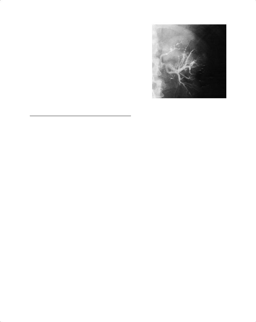
tutional illness, which is often associated with rash, mononeuritis multiplex, vascular hypertension and organ infarction. Polyarteritis nodosa may be confined to the skin. Angiography shows typical microaneurysms (Figure 23.1). Polyarteritis nodosa is associated with hepatitis B infection.
Kawasaki disease (mucocutaneous lymph node syndrome)
An acute vasculitis that primarily affects infants and young children. It presents with fever, rash, lymphadenopathy and palmoplantar erythema. Coronary arteries become affected in up to one-quarter of untreated patients; this can lead to myocardial ischaemia and infarction.
Mediumand small-vessel vasculitis
This group includes the major necrotizing vasculitides: microscopic polyangiitis, Wegener’s granulomatosis and Churg–Strauss syndrome, with involvement of both medium and small arteries. These may occur at any age, with the peak incidence at 60–70 years and are slightly more common in men. The annual incidence is about 20 cases per million people. The symptoms depend on the size and site of the vessel affected and on the individual diagnosis. They are associated with the presence of anti-neutrophil cytoplasmic antibodies (ANCA).
Wegener’s granulomatosis
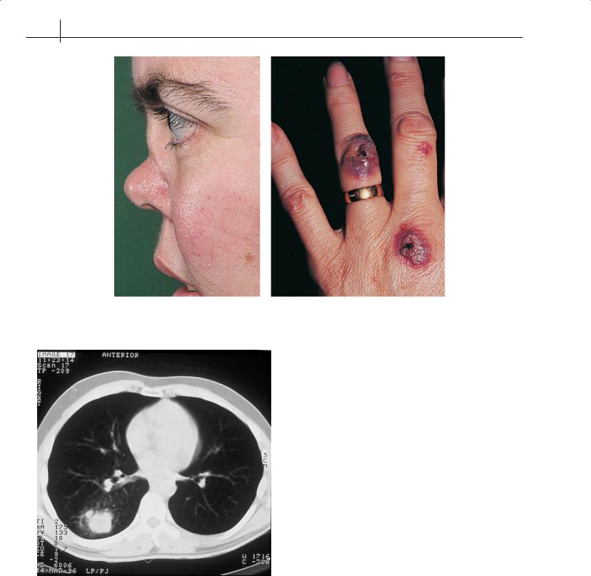
This is characterized by a granulomatous vasculitis of the upper and lower respiratory tracts and glomerulonephritis, but almost any organ system can be affected (Figure 23.2). The lungs are affected in 45% of patients at diagnosis. Symptoms in the ear, nose and throat (such as epistaxis, crusting and deafness) are particularly associated with this condition, and they should be sought in all patients with suspected vasculitis. Patients with limited Wegener’s
granulomatosis—disease without renal involvement—may have a better prognosis. Biopsy of affected organs shows a necrotizing arteritis, often with formation of granulomas (Figure 23.3).
Microscopic polyangiitis
This is characterized by a vasculitis that commonly affects the kidneys. Lung involvement usually presents with haemoptysis caused by pulmonary capillaritis and haemorrhage (pulmonaryrenal syndrome).
Biopsy of the kidney shows a focal segmental necrotizing glomerulonephritis with few immune deposits (sometimes called pauci-immune vasculitis).
Churg–Strauss syndrome
This syndrome is characterized by atopy (especially late-onset asthma), pulmonary involvement (75% of patients have radiographic evidence of infiltration) and eosinophilia in the tissues and peripheral blood (>1 × 10⁹/l). Such features can develop several years before the onset of systemic vasculitis. Cardiac involvement is a particular feature of Churg–Strauss syndrome and determines prognosis. Neuropathy is common.
Small-vessel vasculitis
Small-vessel vasculitis (leucocytoclastic or hypersensitivity) is usually confined to the skin, but it may be part of a systemic illness. The rash is purpuric, sometimes palpable, and occurs in dependent areas. The lesions may become bullous and ulcerate. Nailfold infarcts occur. Biopsy shows a cellular infiltrate of small vessels often with leucocytoclasis (fragmented polymorphonuclear cells and nuclear dust). Small-vessel vasculitis has a number of causes, of which drugs and infection are the most common.
150 ABC of Rheumatology
(a) |
(b) |
Figure 23.2 Wegener’s granulomatosis; (a) typical saddle nose deformity (reproduced with patient’s permission); (b) vasculitic rash
Figure 23.3 Computed tomography scan of thorax, showing a granuloma in Wegener’s granulomatosis
Henoch–Schönlein purpura
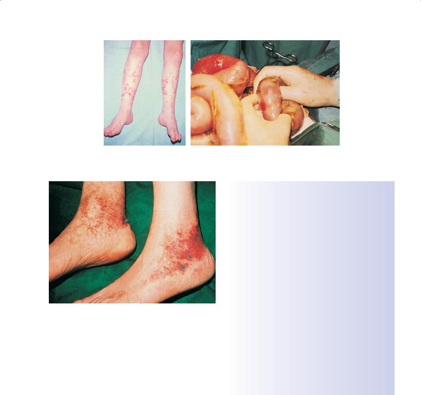
This is a form of small-vessel vasculitis that occurs mainly in children and young adults. Patients present with rash, arthritis, abdominal pain and, sometimes, renal involvement (Figure 23.4). Deposits of immunoglobulin A can be detected histologically in the skin and renal mesangium.
Cryoglobulinaemia
Cryoglobulins are plasma proteins that precipitate in the cold. The condition presents with rash (including purpura digital ischaemia and ulcers) (Figure 23.5), arthralgia and neuropathy. A strong link exists between infection with hepatitis C virus and essential mixed cryoglobulinaemia: 80–90% of such patients are positive for antihepatitis C virus antibodies.
Behçet’s syndrome
Behçet’s syndrome is a systemic vasculitis of unknown aetiology, characterized by oro-genital ulceration. It is most common in Turkey and Japan. Ocular involvement occurs early in the disease course and affects 50% of patients. The pathergy phenomenon is characteristic and is a non-specific hyperreactivity in response to minor trauma.
Investigation
Investigation aims to establish and confirm the diagnosis, the extent and severity of organ involvement, and disease activity (Box 23.2).
Urine analysis
This is the most important investigation, because the severity of renal involvement is one of the key determinants of prognosis. Detection of proteinuria or haematuria in a patient with systemic illness needs immediate further investigation, and the patient is a medical emergency.
Vasculitis and Related Rashes |
151 |
|
|
|
|
(a) |
(b) |
Figure 23.4 Small-vessel vasculitis in Henoch–Schönlein purpura; (a) affecting the skin; (b) affecting the gut |
|
|
Box 23.2 Investigation of vasculitis |
||
|
Assessing inflammation |
||
|
• |
Blood count and differential (total white cell count, eosinophils) |
|
|
• |
Acute-phase response (erythrocyte sedimentation rate, C-reactive |
|
|
|
protein) |
|
|
• |
Liver function |
|
|
Assessment of organ involvement |
||
|
• |
Urine analysis (proteinuria, haematuria, protein excretion) |
|
|
• |
Renal function (creatinine clearance, 24-hour protein excretion, |
|
|
|
urine protein/creatinine ratio biopsy) |
|
|
• Chest radiograph |
||
|
• |
Liver function |
|
|
• |
Nervous system (nerve-conduction studies, biopsy) |
|
|
• |
Cardiac function (electrocardiography, echocardiography) |
|
|
• Gut (angiography) |
||
Figure 23.5 Vasculitic rash in cryoglobulinaemia |
Immunological tests |
||
• |
Antineutrophil cytoplasmic antibodies (including proteinase 3 |
||
|
|||
|
|
and myeloperoxidase antibodies) |
|
|
• |
Other autoantibodies (rheumatoid factor, antinuclear antibodies, |
|
Blood tests |
|
anticardiolipin antibodies) |
|
• |
Complement |
||
Leucocytosis suggests a primary vasculitis or infection. Leucopaenia |
|||
• |
Cryoglobulins |
||
is associated with vasculitis secondary to a connective tissue disease |
|||
Differential diagnosis |
|||
(typically systemic lupus erythematosus). Eosinophilia suggests |
|||
• |
Blood cultures |
||
Churg–Strauss syndrome or a drug reaction. |
|||
• |
Viral serology |
||
|
|||
Liver function tests |
• |
Echocardiography |
|
|
|
||
Abnormal results suggest viral infection (hepatitis A, B or C) or may be non-specific.
Immunology
ANCA are associated with the primary systemic necrotizing vasculitides. ANCA in association with proteinase 3 antibodies are highly specific (>90%) for Wegener’s granulomatosis. Perinuclear ANCA (pANCA) associated with myeloperoxidase antibodies occur in microscopicpolyangiitisandChurg–Strausssyndrome.Rheumatoid factors and antinuclear antibodies may indicate vasculitis associated with connective tissue disease. Complement levels are low in infection, lupus and cryoglobulinaemia.
Biopsy
Tissue biopsy is important to confirm the diagnosis before treatment with potentially toxic immunosuppressive drugs. The choice of tissue to biopsy is crucial.
Other investigations
Angiography can show aneurysms. Blood cultures, viral serology and echocardiography are important to exclude infection and other conditions that may present as systemic multi-system disease and mimic vasculitis (Boxes 23.3 and 23.4).