Материал: ABC-of-Rheumatology-ABC-Series-
152 ABC of Rheumatology
Box 23.3 Important mimics of vasculitis
•Subacute bacterial endocarditis
•Atrial myxoma
•Cholesterol embolism
•Antiphospholipid antibody syndrome
•Calciphylaxis
•Cocaine abuse
Box 23.4 Differential diagnosis of rash and arthritis
•Infection
•Drug reaction
•Sarcoidosis
•Juvenile idiopathic arthritis
•Connective tissue disease
•Psoriasis
•Vasculitis
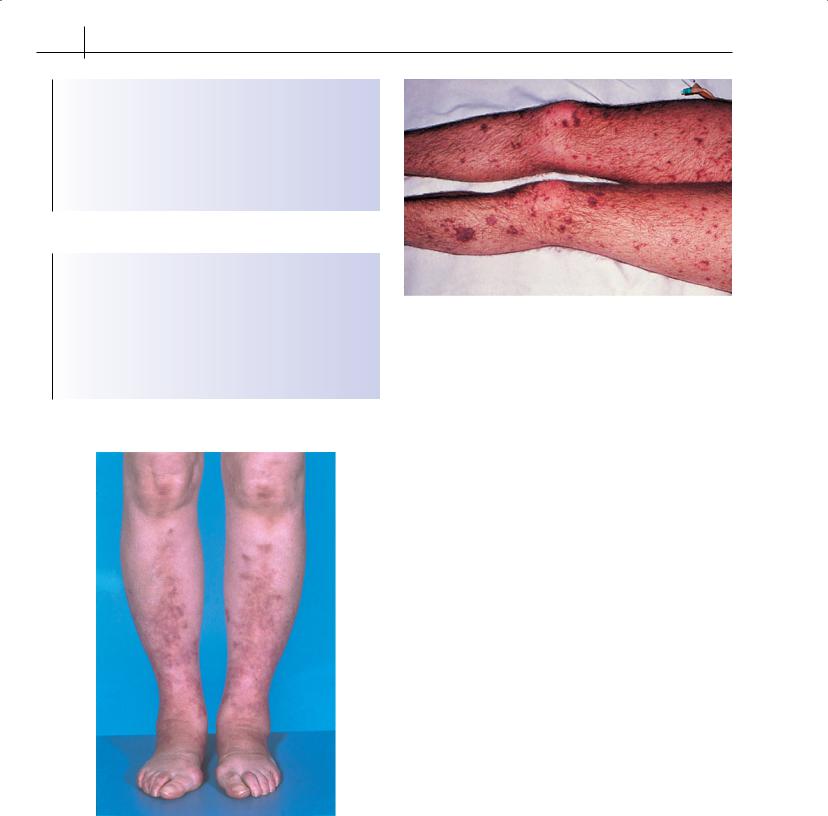
Figure 23.6 Livedo rash in cutaneous polyarteritis nodosa
Differential diagnosis
Livedo reticularis
Livedo reticularis is characterized by persistent patchy reddish-blue mottling of the legs (and occasionally arms) that is exacerbated by
Figure 23.7 Haemorrhagic pustular rash in disseminated infection with
Neisseria meningitidis
cold weather. It may lead to ulceration and is associated with vascular thrombosis (Sneddon’s syndrome) and the presence of antiphospholipid antibodies. It is also a feature of polyarteritis nodosa (Figure 23.6) and cryoglobulinaemia.
Bacterial infections
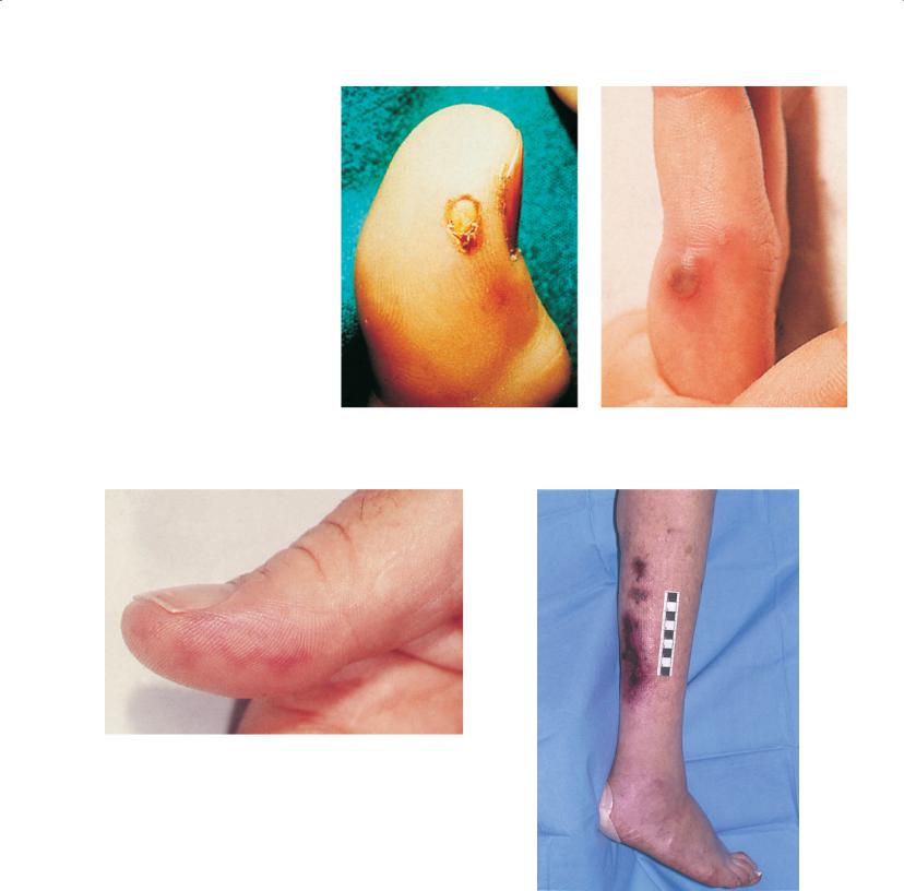
Direct bacterial infection of small arteries and arterioles causes a necrotizing vasculitis or thrombosis. Neisseria meningitidis (Figure 23.7), N. gonorrhoeae (Figure 23.8) and Streptobacillus moniliformis, for example, may infect the vascular endothelium directly and cause maculopapular or purpuric skin lesions. Biopsies of early lesions show small-vessel vasculitis. The organisms can be cultured from an aspirate of the lesions.
Infective endocarditis
Several organisms—streptococci, staphylococci, Gram-negative bacilli and Coxiella—can cause endocarditis. Polyarthritis may be accompanied by splinter haemorrhages, Janeway’s lesions (red macules over thenar and hypothenar eminences) (Figure 23.9), Osler’s nodes (tender papules over extremities of fingers and toes) and clubbing. Diagnosis is by blood culture and echocardiography.
Cholesterol embolism
Cholesterol embolism (Figure 23.10) may occur spontaneously or after trauma to the aortic wall during vascular surgery or angiographic procedures. Typical cutaneous manifestations are ischaemia of the digits, particularly the toes, from abdominal atheroma, emboli and livedo reticularis. Digital ischaemia usually presents as sudden onset of a small, cool, cyanotic and painful area of the foot (usually the toe). The lesions are tender to touch and may progress to ulceration, digital infarction and gangrene; this mimics systemic vasculitis. Presentation may be with a systemic illness caused by tissue inflammation; features include eosinophilia and a positive test for ANCA.
Vasculitis and Related Rashes |
153 |
|
|
|
|
Figure 23.8 Gonococcal pustules in disseminated infection with Neisseria gonorrhoeae
Figure 23.9 Janeway’s lesions in infective endocarditis
Atrial myxoma
Cardiac myxomata are rare benign tumours found most often in the left atrium (90% of cases). Constitutional symptoms and systemic embolization may lead to a wrong diagnosis of vasculitis. Systemic manifestations seen in 90% of cases include fever, weight loss, Raynaud’s phenomenon, clubbing, elevated acute-phase proteins and hypergammaglobulinaemia. It is treated by surgical resection of the primary tumour and emboli.
Antiphospholipid antibody syndrome
Antiphospholipid antibody syndrome may present as catastrophic widespread thrombosis, and this can mimic systemic vasculitis. Livedo reticularis is the most typical cutaneous lesion, and it occurs in association with thrombosis and recurrent fetal loss.
Figure 23.10 Cholesterol emboli
Cocaine abuse
Cocaine abuse can cause destruction of the nasal mucosa and septum, mimicking systemic vasculitis.

154 ABC of Rheumatology
Prognosis
The natural history of untreated primary systemic vasculitis is of a rapidly progressive, usually fatal disease. Before corticosteroids were introduced in Wegener’s granulomatosis, the median survival was 5 months, with 82% of patients dying within 1 year and more than 90% within 2 years. The introduction of corticosteroids improved survival in polyarteritis nodosa to 50% at 5 years. The median survival in Wegener’s granulomatosis was only 12.5 months using corticosteroids alone, with most patients dying of sepsis or uncontrolled disease. The introduction of oral low-dose cyclophosphamide combined with prednisolone resulted in a significant improvement in the mortality of Wegener’s granulomatosis, with a survival rate at 5 years of 82%.
Small-vessel vasculitis confined to the skin without necrotizing features has an excellent prognosis. Takayasu’s arteritis has a good prognosis (3% mortality) but typically relapses.
Treatment
Treatment depends on the size of vessel involved (Box 23.5). Small vessel vasculitis can often be treated conservatively. Takayasu’s arteritis requires high-dose corticosteroids (oral prednisolone 40– 60 mg/day), and additional immunosuppression with methotrexate or azathioprine. The dose of corticosteroid should be reduced rapidly according to clinical and laboratory parameters.
Box 23.5 Aims of management of vasculitis
•Induction of remission
•Maintenance of remission
•Recognition and early treatment of relapse
•Avoidance of drug toxicity
Table 23.2 Treatment regimens for cyclophosphamide
Drug |
Dose |
|
|
Continuous low oral dose |
|
Cyclophosphamide |
2 mg/kg/day |
Prednisolone |
1 mg/kg/day |
Intravenous pulse* |
|
Cyclophosphamide |
10–15 mg/kg† |
Prednisolone |
1 g |
Cyclophosphamide dose should be adjusted according to white cell count, renal function and clinical response
*Pulse frequency: fortnightly (×3), then three-weekly; adjusted according to clinical response and toxicity
†White cell count should be checked 7, 10 and 14 days after the first two pulses and immediately before subsequent pulses. For oral cylophosphamide the white cell count should be checked weekly for one month, fortnightly for two months and then every month.
The recently completed European randomized controlled trials in ANCA-associated vasculitis now guide the treatment approach. For patients with generalized disease, cyclophosphamide (Table 23.2) is used for remission induction and can be given either as continuous low-dose oral therapy or intermittent pulse therapy. Both routes are equally effective at inducing remission, but pulse therapy is probably associated with a slightly higher relapse rate. The major toxicities of cyclophosphamide are haemorrhagic cystitis, formation of bladder tumours, infertility and infection. Toxicity depends on the cumulative dose, so pulse therapy is less toxic. Mesna may reduce the frequency of bladder toxicity with intravenous cyclophosphamide. The risk of ovarian failure depends on age and cumulative dose of cyclophosphamide. Fertile males should be offered sperm storage before they are given cyclophosphamide. Prophylaxis with co-trimoxazole should be considered to prevent infection with Pneumocystis jiroveci. Immunosuppressed patients should receive vaccination with influenza and polyvalent pneumococcal vaccination.
Corticosteroids are started at a dose of 1 mg/kg, and the dose is reduced quite rapidly so that the drug can be discontinued at around 12 months. Alternate-day dosing may reduce the risk of infection. Intravenous methylprednisolone is often given with the first two pulses.
Once remission has been achieved with cyclophosphamide (usually after 3–6 months), azathioprine (or weekly oral methotrexate) is substituted for maintenance therapy. Cyclophosphamide should not be continued for more than 1 year because of the risks of toxicity. Survival has improved and remission can be obtained in most patients (85%) with cyclophosphamide, but many need prolonged immunosuppressive therapy (5–10 years), and the rate of relapse is still substantial (50% at 5 years).
Methotrexate may be considered in patients with localized disease, as an alternative to cyclophosphamide.
Patients with life-threatening disease (pulmonary haemorrhage) or a creatinine >500 μmol/l should receive plasma exchange in addition to intravenous methylprednisolone.
Regular assessment of disease activity is required, and treatment is tailored accordingly. Minor relapses may require an increase in maintenance therapy. Major relapses will require a further course of cyclophosphamide.
Intravenous immunoglobulin is effective in the treatment of Kawasaki disease, but its role in other vasculitides, where it induces temporary improvement, remains controversial at present. Etanercept does not improve relapse rate when used as adjunctive therapy to conventional therapy for remission maintenance. The role of tumour-necrosis-factor- -blocking drugs in induction is uncertain. B-cell depletion with rituximab is a promising approach that is being investigated.
Further reading
Ball GV, Bridges L, eds. Vasculitis, 2nd edn. Oxford: Oxford University Press, Oxford, 2007.
Birck R, Scmitt W, Kaelsch IA, van Der Woude FJ. Serial ANCA determinations for monitoring disease activity in patients with ANCA-associated vasculitis: systematic review. American Journal of Kidney Diseases 2006; 47: 15–23.

Vasculitis and Related Rashes |
155 |
|
|
|
|
De Groot K, Rasmussen N, Bacon P et al. Randomised trial of cyclophosphamide versus methotrexate for induction of remission in early systemic antineutrophil cytoplasmic antibody associated vasculitis. Arthritis and Rheumatism 2005; 52: 2462–2468.
Jayne D, Rasmussen N, Andrassy K et al. A randomised trial of maintenance therapy for vasculitis associated with antineutrophil cytoplasmic auto antibodies. New England Journal of Medicine 2003; 349: 36–44.
Jayne D, Gaskin G, Rasmussen N et al. Randomised trial of plasma exchange or high dose methylprednisolone as adjunctive therapy for severe renal vasculitis. Journal of the American Society of Nephrology 2007; 18: 2180–2188.
Lapraik C, Watts RA, Scott DG. BSR & BHPR guidelines for the management of adults with ANCA associated vasculitis. Rheumatology 2007; 46: 1615–1616.
Maksimowicz-McKinnon K, Clark TM, Hoffman GC. Limitations of therapy and a guarded prognosis in an American cohort of Takayasu arteritis patients. Arthritis and Rheumatism 2007; 56: 1000–1009.
Mukhtyar C, Guillevin L, Dasgupta B et al. EULAR recommendations for the management of primary small and medium vessel vasculitis. Annals of the Rheumatic Diseases 2009; 68: 310–317.
Mukhtyar C, Guillevin L, Cid M et al. EULAR recommendations for the management of large vessel vasculitis. Annals of the Rheumatic Diseases 2009; 68: 318–323.
Reinhold-Keller E, Beuge N, Latza U et al. An interdisciplinary approach to the care of patients with Wegener’s granulomatosis. Arthritis and Rheumatism 2000; 43: 1021–1032.

CHAPTER 24
Laboratory Tests
Cynthia Aranow1, Margaret J Larché2 and David A Isenberg3
1Feinstein Institute for Medical Research, Manhasset, USA
2McMaster University, Hamilton, Ontario, Canada
3University College Hospital, London, UK
OVERVIEW
•Abnormal laboratory tests occur frequently in patients with rheumatologic disorders.
•Laboratory abnormalities suggesting non-specific inflammation are common and accompany many rheumatologic disorders.
•Routine blood tests (haematology and chemistry) are useful to monitor known rheumatologic diseases and may be helpful in diagnosis. Abnormalities may reflect adverse effects of medications or may indicate organ involvement from an underlying rheumatologic disease.
•Immunological testing is primarily for diagnostic purposes and may help subsetting patients (e.g. patients with systemic lupus erythematosus who are anti-Ro positive are more likely to be photosensitive); however, the antinuclear antibody test is not a diagnostic test.
•Genetic testing (such as HLA-B27) is expensive and not a diagnostic tool.
This chapter describes investigations that may be performed in a patient with suspected and known rheumatologic disorders. Abnormal haematology tests, particularly anaemias and platelet abnormalities, are found commonly. Biochemical abnormalities include raised protein and globulin levels and reflect a non-specific inflammatory response. Haematological and biochemical investigations are useful for both diagnostic and monitoring purposes, while most immunological investigations are mainly used to facilitate diagnosis.
Haematology investigations
A full blood count and erythrocyte sedimentation rate (ESR) are used to monitor disease activity, to assess the effects of drug treatment, to exclude factors such as dietary deficiency or haemolysis that may be contributing to the morbidity of a rheumatological
ABC of Rheumatology, 4th edn. Edited by Ade Adebajo. ©2010 Blackwell Publishing Ltd. 9781405170680.
disease, and (rarely) to exclude a primary haematologic malignancy that can mimic various forms of arthritis (Table 24.1).
Platelet abnormalities
Platelet abnormalities are often seen in rheumatic disorders; the most common abnormality is a mild to moderate thrombocytosis, which correlates with disease activity. Thrombocytopenia may occur as a side effect of interventional treatments such as methotrexate, cyclophosphamide or mycophenolate mofetil. Thrombocytopenia may also be observed in patients receiving treatment with gold or penicillamine; however, these medications are now rarely used. An autoimmune thrombocytopenia (usually chronic but occasionally acute) occurs in up to 20% of patients with lupus and in patients with primary antiphospholipid antibody syndrome. In some of these patients it has been possible to demonstrate the presence of antiplatelet antibodies. Approximately 15% of patients with “idiopathic” thrombocytopenia later develop lupus. Thrombocytopenia may also be seen in the subset of rheumatoid arthritis with Felty’s syndrome (see below). Infections associated with arthralgia such as cytomegalovirus, hepatitis C and HIV can additionally be associated with thrombocytopenia.
White blood cell abnormalities
Felty’s syndrome, the association of rheumatoid arthritis with leucopenia (predominantly neutropenia) and splenomegaly (and often leg ulcers), is rare. Leucopenia, particularly lymphopenia, is common in lupus. Bone-marrow suppression is a well-recognized complication of immunosuppressive drugs such as azathioprine, methotrexate, leflunomide, sulfasalazine, cyclophosphamide and mycophenolate mofetil, which are used to treat rheumatoid arthritis, psoriatic arthritis and lupus. Patients taking these drugs require regular haematological assessments to allow early detection of bone-marrow suppression. Leucocytosis is occasionally found in flares of lupus, but is more often a reflection of corticosteroidinduced demargination of neutrophils. Infective causes of a leucocytosis (particularly neutrophilia) must be excluded. Less common abnormalities, such as monocytopenia and eosinophilia in rheumatoid arthritis and basopenia in lupus, are well described. A range of blood test abnormalities in rheumatological disease are shown in Table 24.2.
156