Материал: ABC-of-Rheumatology-ABC-Series-

Raynaud’s Phenomenon and Scleroderma |
127 |
|
|
|
|
Box 19.4 Principles of management of systemic sclerosis
•Accurate diagnosis
•Appropriate subsetting
•Staging disease within subset
•Vascular and immunosuppressive therapy according to stage and subset
•Risk stratification for major organ-based complications based upon serological, genetic and clinical features
•Screening and early intervention when complications develop
•Organ-based therapy: gastrointestinal, respiratory, cardiac, renal
pulmonary or renal complications to be identified. In the future, such information is likely to direct management and screening. Antibodies can predict particular complications such as antitopoisomerase and lung fibrosis, anti-RNA polymerase and renal crisis or anti-Th/To and respiratory involvement in lcSSc. At present the strongest genetic associations relate to SSc-specific autoantibodies, but other genetic factors that determine the disease profile are being sought. Recently a genetic variant in connective tissue growth factor (CTGF) was associated with SSc (Fonseca et al., 2007).
Management of SSc
The principles of effective management of SSc are summarized in Box 19.4. Unfortunately at present there are no disease-modifying treatments of proven efficacy. Most patients benefit from vascular therapy, and a number of agents that suggest the potential for vascular remodelling have been used in trials (Table 19.3). Immunosuppressive treatment is generally reserved for patients with early and aggressive dcSSc or with a major organ-based complication such as interstitial lung disease or myositis. Cyclophosphamide has been demonstrated to have modest benefit in prospective clinical trials (Hoyles et al., 2006; Tashkin et al., 2007). A number of approaches are being evaluated in clinical trials. High-dose immunosuppression with autologous peripheral stem-cell rescue is currently being evaluated in clinical trials. There are currently no effective antifibrotic agents for established SSc, but a number are under development. They include biological therapies that neutralize key potential cytokines driving SSc, including transforming growth factor beta 1 (TGFβ1) and CTGF.
Organ-based complications
The outcome of SSc is largely determined by the extent and severity of organ-based complications. Some of these are almost universal, such as oesophageal reflux, while many of the severe complications occur in around 10–15% of cases overall.
Pulmonary hypertension—Pulmonary hypertension is the single largest cause of death directly attributable to SSc. The frequency of the complication is likely to be around 10% overall, although published prevalence studies have varied largely owing to differences in diagnostic methods and variation in study cohorts. It occurs in both limited and diffuse cutaneous subsets, although as an isolated
Table 19.3 Drugs to treat Raynaud’s phenomenon
Pharmaco-nutrients
Antioxidant vitamins: vitamins C and E at high dose may reduce oxidant stress in Raynaud’s and SSc
Fish oils: maxepa, cod liver oil supplements may favour endothelial production of vasodilatory prostanoids
Gamolenic acid: mechanism uncertain
Seredrin: Gingko biloba, vasodilator giving symptomatic benefit in Raynaud’s
Vasodilators
Calcium channel blockers: nifedipine and others may be effective; primary Raynaud’s responds better than secondary; different agents may demonstrate varied effectiveness and side effects; side effects are frequent and often dose-limiting, including headache, ankle swelling and postural hypotension
Angiotensin receptor blockers: effective for SSc-associated hypertension; may also help Raynaud’s symptoms and have potential beneficial effect on vascular remodelling
Nitroglycerin: topical glyceryl trinitrate ointment or patches may benefit ischaemic digits, but limited by side effects; new microemulsions under evaluation with better tolerability
Other agents
5HT reuptake inhibitors: give well-tolerated and effective symptom relief in many Raynaud’s patients; mechanism of action probably involves depletion of platelet serotonin
Surgical options include lumbar sympathectomy for lower limb symptoms and digital sympathectomy (adventectomy) for severely affected digits; cervical sympathectomy disappointing and only temporarily helpful
Lifestyle adjustments (especially cold avoidance and cessation of smoking), silk-lined gloves and chemical hand-warmers often provide substantial benefit
complication it is most often seen in established lcSSc. Diagnosis can only be made robustly using right-heart catheterization and is determined by a mean pulmonary arterial pressure in excess of 25 mm Hg at rest, without elevated pulmonary capillary wedge pressure, but with increased pulmonary vascular resistance.
Distinction should be made between pulmonary arterial hypertension (PAH) and pulmonary hypertension secondary to severe interstitial lung fibrosis. The latter is uncommon in SSc, although some degree of lung fibrosis in association with PAH is frequent and may impact on survival. Historically the outcome in SSc-associated PAH was dismal, with a 2-year survival of 47%. With the advent of modern advanced therapies, including endothelin receptor antagonists (e.g. bosentan, sitaxentan) and selective phosphodiesterase inhibitors that promote nitric-oxide- induced vasodilatation (e.g. sildenafil) given in the context of a multidisciplinary pulmonary hypertension service, outcome for confirmed PAH has improved and currently survival of greater that 69% at 2 years has been demonstrated. Nevertheless, this is much inferior to outcome in idiopathic (previously primary) PAH using the same therapies, and so there is much scope for improvement.
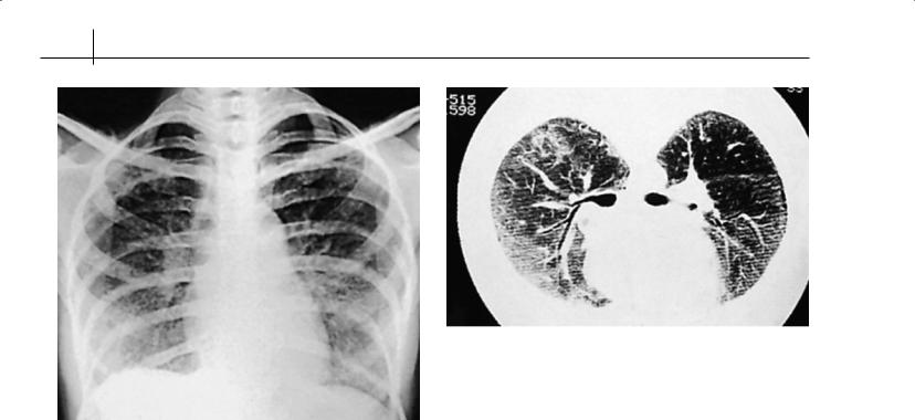
128 ABC of Rheumatology
Figure 19.4 Chest radiograph of diffuse interstitial lung disease in a patient with scleroderma
In SSc there is the potential for earlier diagnosis through screening, and currently it is recommended that all cases of SSc have annual assessment by echo-Doppler and lung function tests. Elevation of estimated right ventricular systolic pressure based on tricuspid regurgitant jet velocity (above 35 mm Hg) or fall in carbon monoxide transfer factor (DLCO) below 55% predicted with preserved lung volumes should prompt consideration for right-heart catheter study. Less invasive tests are being developed, including plasma N-terminal pro-BNP, which has been shown to predict outcome and associate with haemodynamic variables in PAH. In the future is likely that earlier detection and intervention, possibly using combination therapy including ERA, PDE5 inhibition and prostacyclin analogues, will further improve outcome in this difficult problem.
Lung fibrosis—Interstitial lung fibrosis is a common internal organ manifestation of SSc (Figures 19.4 and 19.5). It is present in more than 30% of cases but may not be inexorably progressive. The antiscl70 autoantibody provides a useful clinical marker and is generally associated with lung fibrosis in both SSc subsets.High-resolution computed tomography (HRCT) imaging is currently the goldstandard test to detect and determine the pattern and extent of disease. Bronchoalveolar lavage is favoured in some centres and certainly correlates with the extent of disease, but not always with activity. Lung biopsy is not routinely indicated, but histologically most cases of SSc-associated lung fibrosis are classified as nonspecific interstitial pneumonia, rather than the usual interstitial pneumonia pattern of idiopathic lung fibrosis. This may explain the better outcome for most patients with SSc compared with idiopathic pulmonary fibrosis. Immunosuppressive treatment with cyclophosphamide has recently been shown to be superior to placebo in a large controlled clinical trial, but the effect was modest.
Figure 19.5 High-resolution computed tomography scan showing evidence of early interstitial lung disease
Therefore new and better treatments are needed. It has been suggested that alveolar epithelial damage may be more important than inflammation in driving the fibrosis, and research data confirm that markers of epithelial injury or permeability such as serum KL-6 or DTPA clearance may provide predictive information about future decline in lung function. At present having determined the presence of fibrosis by HRCT, the cornerstone of management is serial pulmonary function testing. Progressive deterioration, even if gradual, is a clinical indicator for active treatment. At present immunosuppressive strategies are used. In severe advanced disease without major co-morbidity, single lung transplantation has been shown to be beneficial.
Scleroderma renal crisis (SRC)—There have been major advances in management of renal disease in SSc. The major problem is one of recognition, and education of both patients and physicians is important. SRC often presents non-specifically with headaches and visual disturbances before encephalopathy, cardiac failure or acute oliguric renal failure develop. Treatment with angiotensinconverting enzyme inhibition is mandatory. Patients should be admitted for blood pressure control and monitoring of renal function. Fifty per cent of cases require dialysis, which is temporary in many individuals. There may be significant recovery in renal function for up to 2 years after a renal crisis, and decisions regarding transplantation should be delayed until that time. There is no evidence that prophylactic administration of ACE inhibitors is helpful in preventing renal crisis or improving outcome, and the cornerstone of management is patient education, vigilant blood pressure monitoring, and avoidance of nephrotoxic drugs or high-dose corticosteroids with prompt initiation or appropriate therapy early in the course of the SRC.
Gut disease—The gastrointestinal tract is the most frequently affected organ in SSc. Up to 90% of patients demonstrate oesophageal dysmotility with reflux, and the proton pump inhibitors have dramatically improved symptomatic disease. Strictures are now relatively rare, although vigilance for Barrett’s metaplasia is required. Midgut disease with bacterial overgrowth may respond

Raynaud’s Phenomenon and Scleroderma |
129 |
|
|
|
|
Box 19.5 Patient support organizations
The Raynaud’s and Scleroderma Association Trust
112 Crewe Road, Alsager, Cheshire ST7 25A Telephone (01270) 872776
www.raynauds.org or www.scleroderma.org.uk
The Scleroderma Society
3 Caple Rd London NW10 8AB Tel: 020 8961 4912
www.sclerodermasociety.co.uk
to broad-spectrum antibiotics, although maintenance treatment may be required. Paradoxically, colonic involvement may lead to severe constipation, and anorectal incontinence is prevalent. It is important that acute abdominal complications of SSc are managed conservatively as far as possible, because major abdominal surgery is poorly tolerated owing to SSc-related co-morbidity, prolonged post-operative ileus and poor healing.
Patient Support organizations are listed in Box 19.5.
References
Fonseca C, Lindahl GE, Ponticos M et al. A polymorphism in the CTGF promoter region associated with systemic sclerosis. New England Journal of Medicine 2007; 357: 1210–1220.
Hoyles RK, Ellis RW, Wellsbury J et al. A multicenter, prospective, randomized, double-blind, placebo-controlled trial of corticosteroids and intravenous cyclophosphamide followed by oral azathioprine for the treatment
of pulmonary fibrosis in scleroderma. Arthritis and Rheumatism 2006; 54: 3962–3970.
Tashkin DP, Elashoff R, Clements PJ; Scleroderma Lung Study Research Group. Effects of 1-year treatment with cyclophosphamide on outcomes at 2 years in scleroderma lung disease. American Journal of Respiratory and Critical Care Medicine 2007; 176: 1026–1034.
Further reading
Akram MR, Handler CE, Williams M et al. Angiographically proven coronary artery disease in scleroderma. Rheumatology (Oxford) 2006; 45: 1395–1398.
Bouros D, Wells AU, Nicholson AG et al. Histopathologic subsets of fibrosing alveolitis in patients with systemic sclerosis and their relationship to outcome. American Journal of Respiratory and Critical Care Medicine 2002;
165: 1581–1586.
Coghlan JG, Mukerjee D. The heart and pulmonary vasculature in scleroderma: clinical features and pathobiology. Current Opinion in Rheumatology 2001; 13: 495–499.
Denton CP, Black CM. Scleroderma and related disorders: therapeutic aspects. Bailliere’s Best Practice & Research. Clinical Rheumatology 2000; 14: 17–35.
LeRoy EC, Medsger TA Jr. Criteria for the classification of early systemic sclerosis. Journal of Rheumatology 2001; 28: 1573–1576.
Silver RM. Clinical aspects of systemic sclerosis (scleroderma). Annals of the Rheumatic Diseases 1991; 50 (Suppl. 4): 854–861.
Steen VD, Medsger TA Jr. Severe organ involvement in systemic sclerosis with diffuse scleroderma. Arthritis and Rheumatism 2000; 43: 2437–2444.
Williams MH, Das C, Handler CE et al. Systemic sclerosis associated pulmonary hypertension: improved survival in the current era. Heart 2006; 92: 926–932.
Williams MH, Handler CE, Akram R et al. Role of N-terminal brain natriuretic peptide (N-TproBNP) in scleroderma-associated pulmonary arterial hypertension. European Heart Journal 2006; 27: 1485–1494.

CHAPTER 20
Reflex Sympathetic Dystrophy
Chris Deighton1 and Paul Davis2
1Derbyshire Royal Infirmary, Derby, UK
2University of Alberta, Edmonton, Canada
OVERVIEW
•Reflex sympathetic dystrophy (RSD) is a descriptive term for a condition mainly affecting the limbs, with severe pain, a preceding event that might be relatively trivial in traumatic terms, and abnormal blood flow and sweating in the affected area.
•Eventual structural changes to superficial and deep structures lead to atrophic, shiny skin, contractures and patchy osteoporosis around joints on X-rays.
•The cause is not clearly understood, but probably a variety of central and peripheral voluntary and involuntary neurological pathways play a part.
•The diagnosis is usually clinical, although X-rays and bone scans may show a characteristic appearance.
•Treatment is empirical, but relies on pain reduction, early mobilization and restoration of function. Further randomized controlled trials are needed to improve the therapeutics of this difficult condition.
Introduction
Reflex sympathetic (osteo)dystrophy (RSD) is a descriptive term for a poorly understood clinical condition of unknown aetiology. It has also been variously termed shoulder-hand syndrome, Sudeck’s atrophy and algodystrophy. It has often been confused and compared with causalgia, a different condition with similar clinical symptoms. Generally speaking, the more words used in the description of a condition, the less we understand that condition. In 1993, it was suggested that reflex sympathetic dystrophy be renamed “complex regional pain syndrome (CRPS) type I”. This change in nomenclature has done little to reassure the nonspecialist that our understanding of the condition has substantially
ABC of Rheumatology, 4th edn. Edited by Ade Adebajo. ©2010 Blackwell Publishing Ltd. 9781405170680.
improved. The change in terminology has also failed to catch on, so that many specialists still refer to RSD, even though it is clear that the reflexes are not necessarily involved, and the sympathetic nervous system cannot be implicated in many patients (for example, sympathetic ganglia blockade only relieves the pain in some patients).
To have the “full house” clinically, the following should be present: (a) severe pain, usually starting peripherally, and working more proximally over time in a non-dermatomal fashion (allodynia)—the pain is disproportionate to the triggering event and clinical findings (hyperpathia); (b) usually a preceding event that might be relatively trivial in traumatic terms; (c) abnormal blood flow to the affected area (usually a limb), with colour changes (blues, whites and reds) and oedema; (d) abnormal sweating in the area; (e) changes in the motor system, with weakness and sometimes tremor; (f) eventual structural changes to superficial and deep structures leading to atrophic, shiny skin, contractures and patchy osteoporosis around joints on X-rays.
Although diagnostic criteria have been proposed, these have not been validated and are complicated by the fact that not all features may be present at the same time and may vary in their intensity. The condition tends to affect upper limbs more commonly than lower limbs. Usually one limb is affected, but it can become bilateral, or affect another limb. It is usually most evident distally (hand and wrist, or foot and ankle), but a whole limb can be affected, such as in “shoulder-hand syndrome”.
This chapter explores the following areas: What causes RSD? How is RSD diagnosed? What is the treatment of RSD?
What causes RSD?
The cause of RSD is far from understood, but it appears to involve an exaggeration of normal physiological responses and involves changes at multiple levels in the central and peripheral nervous systems. Some epidemiological features of RSD are shown in Table 20.1. Taking total knee arthroplasty as an example, a prevalence of between 0.8 and 1.2% of persistent RSD has been quoted. However, a recent prospective study suggested that 21% of patients fulfilled diagnostic criteria 1 month after operation, falling to 12.7% at 6 months, suggesting that symptoms and signs of RSD are not
130

Reflex Sympathetic Dystrophy |
131 |
|
|
|
|
Table 20.1 Some epidemiological factors in reflex sympathetic dystrophy
Triggers |
Usually some noxious event such as: |
|
Wrist and tibial fractures (about 30% may |
|
demonstrate mild features, but only a minority |
|
go on to severe disease) |
|
Trauma: mild or moderate |
|
Rotator cuff tendonitis or subacromial bursitis |
|
Surgery: carpal tunnel decompression, |
|
arthroscopy, arthroplasty, lumbar spine surgery |
|
Central nervous system disorders: head injury, |
|
hemiplegia, spinal cord injury, neuropathy |
|
Myocardial infarction |
|
Immobilization: in any of the above may be an |
|
important factor |
Sex |
More common in women than men, with a ratio |
|
of 3:1 quoted |
Age |
Any age, although the mean in some studies is |
|
quoted as 52 years; now well recognized in |
|
children |
Genetics |
Some evidence to support a familial predisposition |
Personality traits |
No convincing evidence to support an association |
Psychological factors |
Some patients can have motor weakness and |
|
movement disorders relieved by placebo, nerve |
|
blocks or infusions |
|
|
uncommon after operation, but persistent full-blown disease is mercifully unusual. Figures of up to 35% and 5% have been reported for Colles’ fracture and peripheral nerve injury, respectively. The pathology of RSD is bedevilled by the lack of tissue studies, either preor post-mortem. Limited histological investigations have suggested that microangiopathy or other vascular abnormalities may be a key driver. A crucial question that has not been satisfactorily answered is: Why do the majority of patients who suffer the potential triggers listed in Table 20.1 make a full and uneventful recovery, but a minority go onto develop RSD? A number of theories have been propounded, but revolve around peripheral mechanisms, central mechanisms and neurogenic inflammation with microvascular dysfunction. These interrelate in a series of vicious circles that result in the characteristic features of RSD, which are summarized below.
Peripheral mechanisms
Trauma to C fibres and A™ afferents is likely to be an initiating event. Many patients have sympathetically maintained pain, which may activate both mechanoreceptors and nocioceptors. Some patients experience benefit from alpha blockade, supporting a role for -adrenoceptors in the pathogenesis of RSD. These receptors become expressed on nocioceptors in some cases of soft-tissue and nerve injury. Some patients demonstrate supersensitivity to catecholamines, consistent with increased -adrenoceptor responsiveness.
Central mechanisms
In RSD an initial activation of nocioceptors may lead to alteration of central information processes, resulting in central sensitization. Patients exhibit normal thresholds for the detection of cold and heat, but reduced thresholds for cold-pain and heat-pain, suggest-
ing a central nervous disturbance. Activation of low-threshold mechanoreceptors is interpreted as noxious, and results in normal sensations being interpreted as painful (“allodynia”). There is a close similarity between the autonomic features of RSD, and those of autonomic failure after stroke. The latter occur in the absence of pain, suggesting an uncoupling of the mechanisms that underpin the pain and sensory symptoms from the autonomic features. Tests on normal volunteers that create conflict between motorsensory central nervous processing can lead to pain and sensory disturbances, such as using mirrors during congruent and incongruent limb movements. It has been proposed that central processing of persistent sensorymotor conflict may lead to chronic pain in some vulnerable individuals.
Neurogenic inflammation
Release of vasoactive peptides, including substance P and calcitonin gene-related peptide, from afferent nerve fibres cause vasodilatation, with increased vascular permeability and protein leakage. Neuropetides may also be released in response to impaired blood flow, oxygen deficiency and an increase in protons and skin lactate levels. This might explain why some of the early clinical features of RSD appear to be inflammatory.
Microvascular dysfunction
A number of investigators have confirmed microvascular dysfunction in RSD, although it remains unknown whether these changes, reflected by colour and temperature changes, drive the disease process or are secondary to it.
Bringing these factors together, it has been suggested that RSD is initiated by trauma to C fibres and A™ afferents in soft tissue or nerves, resulting in neurogenic inflammation. Signs of inflammation predominate in early disease, with redness, increased skin temperature due to inhibition of cutaneous vasoconstrictor neurons, with subsequent loss of function and pain. Early in the disease, the sympathetic nervous system plays a role, but when central sensitization takes over, with changes at the dorsal root ganglion level, the pain becomes independent of sympathetic nerves. There is a competition between the continued inhibition of vasoconstriction and supersensitivity of the peripheral vessels to circulating adrenaline. Late intractable disease can be characterized by a cold, painful limb with poor or no function, with disuse leading to immobility and contractures.
How is RSD diagnosed?
In the early stages of RSD the limb is swollen and tender, and the diagnosis may not be straightforward, as it can mimic many other diseases, such as inflammatory arthritis, cellulitis, osteomyelitis, deep venous thrombosis, lymphatic obstruction and malignancy. In the late intractable disease, when the limb becomes cold, chronic arterial insufficiency needs to be considered.
There is no diagnostic test for RSD, and tests are only required to rule out the other causes of a painful swollen limb as listed above. Routine investigations, such as a full blood count and erythrocyte sedimentation rate should be normal, and if not an explanation should be sought. In terms of positive features supporting RSD