Материал: ABC-of-Rheumatology-ABC-Series-

132 ABC of Rheumatology
Table 20.2 Diagnostic criteria for RSD
1. The presence of an initiating noxious event, or a cause of immobilization
2. Continuing pain, allodynia or hyperalgesia, in which the pain is disproportionate to any inciting event
3. Evidence at some time of oedema, changes in skin blood flow, or abnormal sudomotor activity in the region of the pain
4. This diagnosis is excluded by the existence of conditions that would otherwise account for the degree of pain and dysfunction
Note: criteria 2–4 must be satisfied
(Table 20.2), X-rays may show patchy osteoporosis, especially in the juxta-articular region. Joint space is usually preserved, but may be lost in late disease with ankylosis. On bone scanning there is increased uptake in early disease, and reduced uptake in late disease. Patients with markedly increased uptake may have a better prognosis, possibly reflecting the fact that they have not yet progressed to late-stage disease. Thermography detects asymmetry in limb surface temperature, but is not widely available. Bone densitometry and magnetic resonance imaging (MRI) may show nonspecific changes, such as reduced bone density, soft-tissue swelling and bone-marrow oedema, but nothing specific to positively diagnose RSD. The greatest value of MRI is to rule out other causes of a painful swollen limb.
How is RSD treated?
Owing to our limited understanding of the aetiology and pathogenesis of RSD, much of the therapeutics is empirical, and what helps one patient may not help others. Because established RSD can be very challenging to treat, emphasis has been placed on prevention where possible and, failing that, early intervention. The main aims are to reduce pain and restore function. Early mobilization following predisposing conditions is important, and graded physiotherapy may be very helpful. A trial in 1999 showed that vitamin C, a powerful antioxidant, may prevent RSD, supporting the growing evidence for the role of oxygen-free radicals in RSD. This needs to be researched further.
Patients who are particularly vulnerable are patients with a previous history of RSD, particularly if they require surgery on the previously affected part. A controlled study found that the risk of RSD could be reduced by a stellate ganglion block after the operation. An uncontrolled study suggested pre-operative calcitonin may prevent recurrence.
Although many experts and committees have recommended physiotherapy, occupational therapy, vocational rehabilitation and behavioural therapy, the evidence base for these is weak or lacking. One study compared physiotherapy and occupational therapy with social work intervention as the control, and showed no differences in the three groups for pain at 12 months, with only small improvements in temperature and global impairment for the intervention arms of the trial. An algorithm of treatment has been proposed by Stanton-Hicks (2002) with a cautious start (heat, massage and
Table 20.3 Treatment modalities for RSD (modified and simplified from Stanton-Hicks, 2002)
Medical |
Rehabilitation |
Psychological |
|
|
|
Medications |
Motivation |
Counselling |
NSAIDs |
Desensitization |
Behaviour modification |
Opioids |
Isometric exercises |
Coping skills |
Tricyclics |
Mobilization |
Relaxation therapy |
Adrenoceptor |
Flexibility |
Hypnosis |
antagonists |
Strength exercises |
|
Corticosteroids |
|
|
Calcitonin |
|
|
Neurologic blocks |
|
|
Sympathetic |
|
|
Regional |
|
|
Epidural |
|
|
Neurostimulation |
|
|
Peripheral |
|
|
Epidural |
|
|
|
|
|
NSAIDs = non-steroidal anti-inflammatory drugs
gentle movement to restore normal sensory processing), then isometric exercises for strengthening, treatment of secondary myofascial pain syndrome, aerobic conditioning, through to complete functional rehabilitation. Because pain can be the main rate-limit- ing factor in rehabilitation, medical and psychological therapies often have to run side by side (Table 20.3).
The mainstay of drug interventions is analgesics and non-ster- oidal anti-inflammatories. Low-dose antidepressants and anticonvulsants are commonly used, but the evidence base is sparse. A systematic review of therapies concluded that the only trial data that consistently demonstrated analgesia was with oral corticosteroids. However, many clinicians have understandable concerns about using steroids for disease that has the potential to become chronic, and where the evidence base for ongoing inflammation driving the disease is limited. A plethora of other drugs have been tried in RSD, which is testimony to the difficulties in treating the condition. Intranasal calcitonin has shown conflicting results. Drugs that do show promise are the bisphosphonates, justified initially on osteoporosis being a significant feature of RSD. A controlled trial of alendronate showed improved bone mineral content of the affected limb, but only small benefits to pain management. By contrast, a trial of intravenous clodronate showed substantial improvements in pain management at 6 months, with highly significant pain reduction compared with placebo.
The role of sympathetic blockade is controversial. For paravertebral blockade, the stellate ganglion for upper limb RSD is blocked with a series of local anaesthetics, depending on response, and the lumbar sympathetic chain for lower limb RSD. Another technique is intravenous blockade, usually with guanethidine. However, a systematic review found this treatment to be ineffective, so its use may decline in future. Continuous blockade of the brachial or lumbar plexus has been advocated with drugs such as morphine, so that whenever the catheter is in place, the patient can take advantage of the pain relief to maximize their rehabilitation.

Reflex Sympathetic Dystrophy |
133 |
|
|
|
|
Intrathecal baclofen proved to be effective for the upper limb dystonias in six out of seven patients, but did not improve pain. Spinal-cord stimulation has been shown to be effective in relieving pain in controlled trials. The procedure is, however, not without risk, as it involves placing an electrode on the dorsal aspect of the spinal cord, and an electric current produces paraesthesias that block the pain in the affected area. However, the average improvement in pain is sustained but not substantial, and functional and quality-of-life benefits have not been demonstrated. This leaves the dilemma of whether invasive and costly interventions that provide modest pain relief are justified. Clearly these concerns and the risks involved mean that patients have to be carefully selected.
Further reading
Amit S, Williams K, Raja SN. Advances in treatment of complex regional pain syndrome: recent insights on a perplexing disease. Current Opinion in Anaesthesiology 2006; 19: 566–572.
Birklein F. Complex regional pain syndrome. Journal of Neurology 2005; 252: 131–138.
Gispen JG. Painful shoulder and the reflex sympathetic dystrophy syndrome. In: Koopman WJ, ed. Arthritis and Allied Conditions, 14th edn. Lippincott Williams & Wilkins, Baltimore, 2001: 2126–2134.
Harden RN, Bruehl SP. Diagnosis of complex regional pain syndrome: signs, symptoms and new empirically derived diagnostic criteria. Clinical Journal of Pain 2006; 22: 415–419.
Herrick A. Reflex sympathetic dystrophy (complex regional pain syndrome type 1). In: Hochberg MC, Silman AJ, Smolen JS, Weinblatt ME, Weisman MH, eds. Rheumatology. Mosby, Edinburgh, 2003.
Merritt WH. The challenge to manage reflex sympathetic dystrophy/complex regional pain syndrome. Clinics in Plastic Surgery 2005; 32: 575–604.
Stanton-Hicks MD, Burton AW, Bruehl SP et al. An updated interdisciplinary clinical pathway for CRPS: report of an expert panel. Pain Practice 2002; 2: 1–16.

CHAPTER 21
Is it a Connective Tissue Disease?
Peter J Maddison1 and Mohammed Tikly2
1Bangor University, Bangor, UK
2University of the Witwatersrand, Johannesburg, South Africa
OVERVIEW
•Raynaud’s phenomenon and sicca symptoms are common presenting features of connective tissue diseases.
•The indirect immunofluorescence test is sufficient as a screening test for antinuclear antibodies, which are the serological hallmark of connective tissue diseases.
•Patients frequently present initially with non-specific features of a connective tissue disease but do not fulfil criteria for any specific disorder.
•Less than one-third of patients with undifferentiated connective tissue evolve clinically to fulfil classification criteria of a defined connective tissue disease.
•Not only is it important to recognize and treat potentially aggressive disease, but also to avoid over-treatment where the diagnosis is unclear or the disease has a potentially benign course.
Diagnosis of connective tissue diseases is often challenging because of the protean clinical features and the fact that no single symptom or sign is pathognomic of a specific connective tissue disease. Classification criteria for the major connective tissue diseases (see Chapters 18 and 19) were developed primarily as a means of standardizing patient populations for clinical research rather than for diagnosis. In practice, they are extremely limited for early diagnosis of connective tissue diseases. In many patients attending connective disease clinics, it is not possible to make definitive diagnosis, especially early in the course of a systemic rheumatic illness. Many, but not all, of these patients eventually fulfil classification criteria for one or more of the major connective tissue disease entities, a process that may take 10–20 years.
A substantial proportion of these patients have non-specific symptoms or signs—most commonly either isolated Raynaud’s phenomenon, or an early inflammatory polyarthritis, or constitutional symptoms of fever, malaise and fatigue. Serological tests often show abnormalities that are suggestive of a connective tissue disease but not sufficiently specific to classify the patient as having one of the major connective tissue diseases. This has prompted the
ABC of Rheumatology, 4th edn. Edited by Ade Adebajo. ©2010 Blackwell Publishing Ltd. 9781405170680.
introduction of additional terms such as “overlap syndrome” and “undifferentiated connective tissue disease.”
From a management perspective, it is important not only to recognize and treat potentially aggressive disease, but also to avoid over-treatment in patients where either the diagnosis is unclear or the disease has a potentially benign course.
Autoantibody profile in diagnosis
Antinuclear antibodies are a hallmark of connective tissue diseases. Serology is of particular value in situations in which clinical expression of the disease is incomplete, when the presence of a particular antinuclear antibody profile can be diagnostic. These antibodies can be found in a variety of clinical settings, however, and their occurrence does not necessarily indicate the presence of any specific disease. It is therefore imperative that requests for antinuclear antibody tests and the interpretation of results thereof be done in the light of the clinical findings.
The indirect immunofluorescence test, using the HEp-2 cell substrate, is the gold standard for detecting antinuclear antibodies. In both systemic lupus erythematosus and scleroderma, antinuclear antibodies can be detected in 95% or more of untreated patients with active disease by this method (Table 21.1). In cases suspected of having either of these diseases, the indirect immunofluorescence test is enough as a screening test for antinuclear antibodies, and it is not cost effective to test automatically for anti-nDNA or other antibody specificities. The individual antinuclear antibody fluorescent patterns are of limited diagnostic utility but may provide guidance to more specific immunological tests. In some instances, a false negative result may occur if either the antigen is outside the nucleus (for example, anti-Jo-1 and anti-ribosomal P-protein antibodies, both often categorized under the umbrella term “antinuclear antibodies”) or if it is present in a form not recognised by a particular autoantibody (for example, when anti-Ro is directed exclusively to determinants on the native Ro molecule not expressed in cultured HEp-2 cells). In such cases, the clinical picture dictates that specific autoantibody assays should be undertaken.
Once antinuclear antibodies have been detected with a screening test, it is important to determine their specificity. This is now part of the standard operating procedure of serology laboratories, but the process is greatly facilitated by the doctor giving sufficient clinical information when antinuclear antibody testing is requested.
134
Is it a Connective Tissue Disease? |
135 |
|
|
|
|
Table 21.1 Antinuclear antibodies in various diseases detected by indirect immunofluorescence
Condition |
Frequency of antinuclear |
|
antibodies (%) |
|
|
Autoimmune rheumatic disease |
|
• Drug-induced lupus |
100 |
• Systemic lupus erythematosus |
98 |
• Systemic sclerosis (scleroderma) |
95 |
• Sjögren’s syndrome |
80 |
• Pauciarticular juvenile idiopathic arthritis |
70 |
• Polymyositis or dermatomyositis |
60 |
• Rheumatoid arthritis |
50 |
Organ-specific autoimmunity |
|
• Primary autoimmune cholangitis |
100 |
• Autoimmune hepatitis |
70 |
• Myasthenia gravis |
50 |
• Autoimmune thyroid disease |
45 |
• Idiopathic pulmonary hypertension |
40 |
Other conditions |
|
• Waldenström’s macroglobulinaemia |
20 |
• Subacute bacterial endocarditis |
20 |
• Infectious mononucleosis |
15 |
• Leprosy |
15 |
• HIV |
10 |
Normal population |
|
• Children |
8 |
• Adults |
15 |
|
|
Box 21.1 Characteristics of undifferentiated connective tissue disease
•Common manifestations
Raynaud’s phenomenon
Arthralgia or myalgia
Rash
Sicca symptoms
Constitutional symptoms (fever, malaise, fatigue)
•One-third evolve into a defined connective tissue disease, usually within 5 years
Specific antinuclear antibody tests are often helpful in stratifying patients into clinical subsets, which may be useful in the further management of specific clinical manifestations and prognostication (Table 21.2). These autoantibodies are usually present from the beginning of the clinical presentation and are detectable throughout the course of the disease. In some instances, such as the anti-nDNA test, autoantibody titres may fluctuate with disease activity. Many serology laboratories these days use commercial kits to detect specific autoantibodies. Although these tests are less labour intensive, they vary in sensitivity, and sometimes produce false-positive results. This is especially the case with the anti-nDNA and anti-Sm assays.
Undifferentiated connective tissue disease
The term “undifferentiated connective tissue disease” was first coined in 1980 by LeRoy and colleagues (LeRoy et al., 1980). This was to counter the concept of mixed connective tissue disease being a distinct disease entity and to point out that many patients designated as having mixed connective tissue disease presented with an early phase of disease that later evolved into one of the “classic” connective tissue diseases, particularly scleroderma. Subsequently, undifferentiated connective tissue disease has been embraced by others and, rather than replacing mixed connective tissue disease, it has been used to describe patients with clinical and laboratory features of connective tissue disease (Box 21.1) who
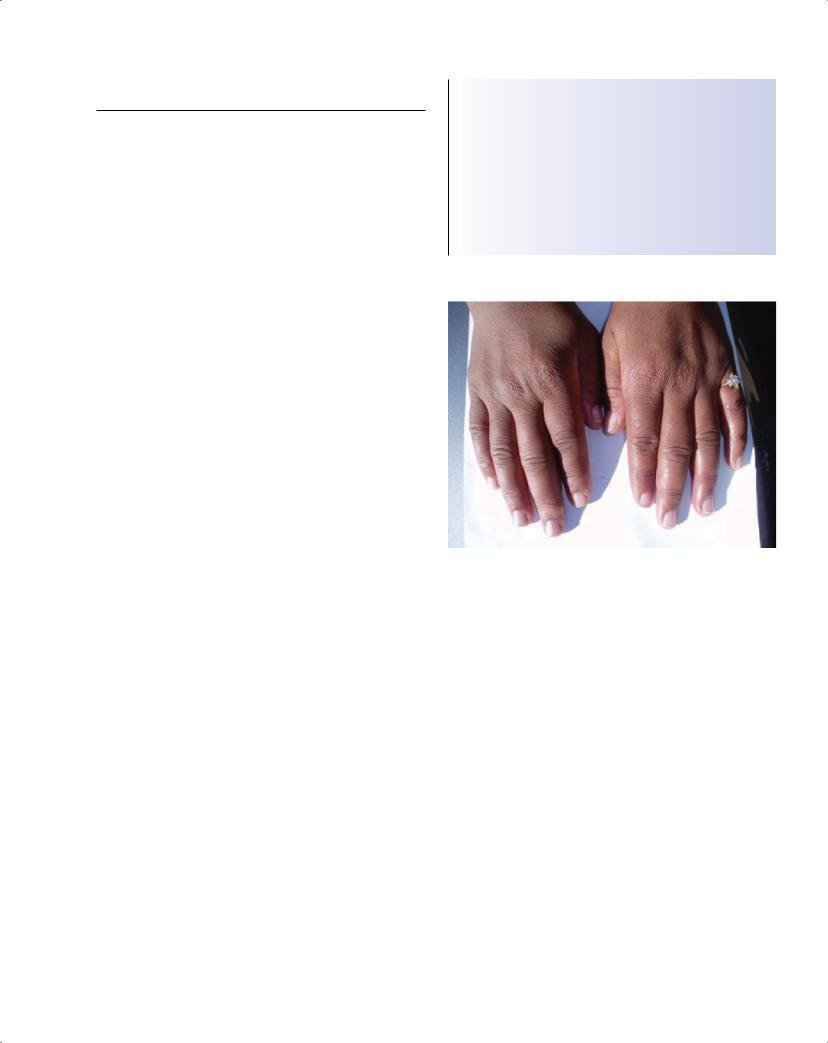
Figure 21.1 Swollen “puffy” fingers of patient with undifferentiated connective tissue disease
do not fulfil criteria for any one disorder (Figure 21.1). Although no universally agreed definition of “undifferentiated connective tissue disease” exists, this term should be distinguished from other commonly used terms such as “overlap syndrome,” in which patients meet the criteria for two or more connective tissue diseases or specific criteria for mixed connective tissue disease (Table 21.3) (see also Chapters 18 and 19). Other terms used in the literature that are synonymous with undifferentiated connective tissue disease include “lupus-like”, “pre-lupus,” “latent lupus” and “incomplete lupus.”
Long-term, prospective follow-up studies of outcome show that these patients represent a large proportion (25–50%) of patients presenting to connective tissue disease clinics. Only a minority of patients, about 30%, evolve clinically to fulfil classification criteria of a defined connective tissue disease, usually in the first few years of follow-up. Spontaneous remission occurs in 5–10% of patients, but in the majority the undifferentiated connective tissue disease state persists. Major organ involvement is rare in these patients. Serositis, alopecia, photosensitivity, discoid rash (Figure 21.2) and the presence of either anti-nDNA antibodies or anti-Sm antibodies are predictors of evolution to SLE.

136 |
|
ABC of Rheumatology |
|
|
|
|
|
|
|
|
|
||
|
|
|
|
|
|
|
|
Table 21.2 Specificity of antinuclear antibodies in diagnosis and disease expression |
|
|
|||
|
|
|
|
|
|
|
|
Disease |
|
Antibody |
Frequency (%) |
Clinical association |
|
|
|
|
|
|
||
Systemic lupus erythematous |
Anti-nDNA‡ |
70 |
• Lupus nephritis |
|||
|
|
|
|
Anti-nucleosome |
70 |
• Early disease, lupus nephritis, |
|
|
|
|
|
|
drug-induced lupus |
|
|
|
|
Anti-Sm |
10–25* |
• Vasculitis, central nervous system |
|
|
|
|
|
|
lupus |
|
|
|
|
Anti-U1RNP |
30–50* |
• Raynaud’s phenomenon, swollen |
|
|
|
|
|
|
fingers, arthritis, myositis, mixed |
|
|
|
|
|
|
connective tissue disease |
|
|
|
|
Anti-Ro |
40 |
• Photosensitive rash, subacute |
|
|
|
|
|
|
cutaneous lupus erythematosus, |
|
|
|
|
|
|
neonatal lupus, congenital heart |
|
|
|
|
|
|
block, Sjögren’s syndrome |
|
|
|
|
Anti-La |
15 |
• As for anti-Ro |
|
|
|
|
Anti-ribosomal P-protein |
15 |
• Central nervous system lupus |
|
|
|
|
|
|
(psychosis or depression) |
|
Sjögren’s syndrome |
Anti-Ro |
60–90# |
• Extraglandular disease, vasculitis, |
||
|
|
|
|
|
|
lymphoma |
|
|
|
|
Anti-La |
35–85# |
• As for anti-Ro |
|
Systemic sclerosis |
Anti-centromere |
5–30† |
• Limited cutaneous disease, |
||
|
|
|
|
|
|
microvascular or macrovascular |
|
|
|
|
|
|
disease, telangiectasia |
|
|
|
|
Anti-ThRNP |
4 |
• Limited cutaneous disease |
|
|
|
|
Anti-topo-1 |
25 |
• Diffuse cutaneous disease, interstitial |
|
|
|
|
|
|
lung disease |
|
|
|
|
Anti-RNA polymerases |
20 |
• Rapidly progressive diffuse cutaneous |
|
|
|
|
|
|
disease, scleroderma renal crisis |
|
|
|
|
Anti-U3RNP |
5–20* |
• Diffuse cutaneous disease, |
|
|
|
|
|
|
pulmonary hypertension |
|
|
|
|
Anti-PM-Scl |
5 |
• Scleroderma–polymyositis overlap |
|
|
|
|
Anti-Ku |
2 |
• Scleroderma–polymyositis overlap |
|
Dermatomyositis and polymyositis |
Anti Jo-1 |
30 |
• Anti-synthetase syndrome: |
||
|
|
|
|
|
|
mechanic’s hands, interstitial lung |
|
|
|
|
|
|
disease |
|
|
|
|
(antibodies to other tRNA synthetases) |
(3) |
(anti-synthetase syndrome) |
|
|
|
|
Anti-SRP |
4 |
• Severe myositis |
|
|
|
|
Anti-Mi2 |
10 |
• Dermatomyositis |
‡Anti-double-stranded DNA antibody
*Higher frequency in people of African or Indian origin
# With sensitive enzyme-linked immunosorbent assays
†Low frequency in people of African origin
Table 21.3 Terminology
Undifferentiated connective |
Patient has features seen in connective |
tissue disease |
tissue disease but does not meet |
|
criteria for a defined connective tissue |
|
disease |
Overlap syndrome |
Patients meet criteria for two or more |
|
connective tissue diseases |
Mixed connective tissue disease |
Overlap of rheumatoid arthritis-like |
|
arthritis, systemic lupus erythematosus, |
|
scleroderma, and myositis with |
|
antibodies to U1RNP |
|
|
Box 21.2 Raynaud’s phenomenon—features suggestive of an underlying connective tissue disease
•Onset in early childhood or later adult life
•Asymmetrical involvement of fingers
•Evidence of digital ischaemic damage
•Abnormal morphology of nailfold capillaries (including dilated, distorted capillaries and areas of capillary dropout)
•Presence of autoantibodies associated with connective tissue disease
Raynaud’s phenomenon
Raynaud’s phenomenon is often the presenting manifestation of connective tissue diseases, especially scleroderma (Box 21.2). It is common, however, in otherwise healthy people.
Which connective tissue disease?
Although clinical presentation in the early stage can be similar between connective tissue diseases, the evolution of typical clinical features over weeks or months is usually enough to distinguish the