Материал: ABC-of-Rheumatology-ABC-Series-

122 ABC of Rheumatology
Bertsias GK, Ioannidis JP, Boletis J et al. EULAR recommendations for the management of systemic lupus erytematosus (SLE): report of a task force of the European Standing Committee for International Clinical Studies Including Therapeutics. Annals of the Rheumatic Diseases 2008; 67: 195–205.
Danchenko N, Satia JA, Anthony MS. Epidemiology of systemic lupus erythematosus: a comparison of worldwide disease burden. Lupus 2006; 15: 308–318.
Derksen RH, Khamashta MA, Branch DW. Management of the obstetric antiphospholipid syndrome. Arthritis and Rheumatism 2004; 50: 1028–1039.
Gayed M, Gordon C. Pregnancy and rheumatic diseases. Rheumatology (Oxford) 2007; 46: 1634–1640.
Hochberg MC. Updating the American College of Rheumatology revised criteria for the classification of systemic lupus erythematosus. Arthritis and Rheumatism 1997; 40: 1725.
Miyakis S, Lockshin MD, Atsumi T et al. International consensus statement on an update of the classification criteria for definite antiphospholipid syndrome (APS). Journal of Thrombosis and Haemostasis 2006; 4: 295–306.
Mok CC, Lau CS. Pathogenesis of systemic lupus erythematosus. Journal of Clinical Pathology 2003; 56: 481–490.
Ostensen M, Khamashta M, Lockshin M et al. Anti-inflammatory and immunosuppressive drugs and reproduction. Arthritis Research and Therapy 2006; 8: 209.
Tan EM, Cohen AS, Fries JF et al. The 1982 revised criteria for the classification of systemic lupus erythematosus. Arthritis and Rheumatism 1982; 25: 1271–1277.

CHAPTER 19
Raynaud’s Phenomenon and Scleroderma
Christopher P Denton and Carol M Black
Royal Free Hospital, London, UK
OVERVIEW
•Survival from systemic sclerosis (SSc) is much better than in the past, owing to better and more effective treatment of organ-based complications, including scleroderma renal crisis and pulmonary arterial hypertension.
•Lung fibrosis is present in approximately one-third of systemic sclerosis patients but may be trivial and not progress. Progressive disease is currently treated with intravenous cyclophosphamide.
•Internal organ manifestations of systemic sclerosis occur in both of the major subsets (limed cutaneous SSc and diffuse cutaneous SSc). Association of anti-topoisomerase 1 with lung fibrosis and anti-RNA polymerase III autoantibodies with renal crisis is seen in both subsets.
•In most cases of dcSSc, peak severity of skin sclerosis is within the first 18 months of disease and the skin often plateaus or improves.
•Approximately 10% of patients with isolated Raynaud’s phenomenon that have positive antinuclear antibodies and abnormal digital nailfold capillaroscopy will later develop clinical features of connective tissue disease.
disease from those in which internal organ complications may develop is central to management. Absence of antinuclear antibodies, altered nailfold capillaroscopy and Raynaud’s phenomenon all point towards localized scleroderma (see below).
Raynaud’s phenomenon
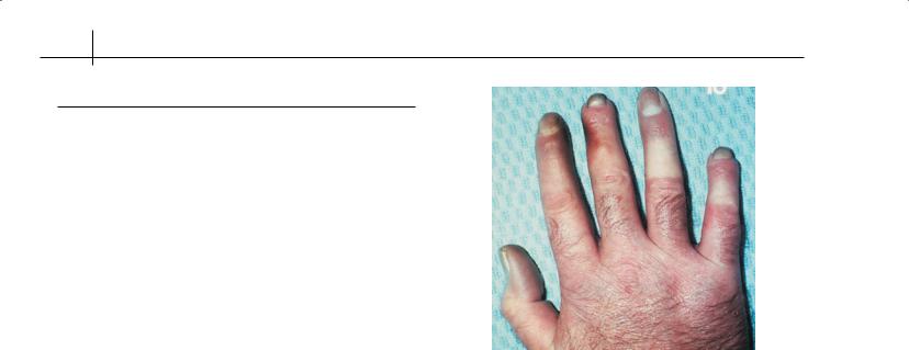
Episodic cold-induced vasospasm (Figure 19.1), triggered by cold or emotional stress, affects around 5% of the adult population, especially young females. In Primary Raynaud’s (90%) there are no other clinical or investigational abnormalities. Secondary Raynaud’s (10%) implies there are other features, usually an underlying autoimmune rheumatic disease. Investigation of Raynaud’s symptoms includes the identification of secondary causes (Box 19.1). Such causes of Raynaud’s, or acrocyanosis, include vibrating machine tools, thoracic-outlet obstruction, drugs such as β- blockers, and haematological abnormalities such as cryoglobulinaemia. Macrovascular arterial disease, embolization and systemic vasculitis, including Berger’s disease, are important but rare differential diagnoses. Some patients with isolated Raynaud’s
The scleroderma spectrum
Scleroderma, meaning “hard skin”, is a generic term used to describe a number of related connective tissue disorders. There is a spectrum from localized dermal sclerosis, through systemic conditions featuring cutaneous and internal organ fibrosis together with vascular dysfunction, to purely vascular disorders of Raynaud’s phenomenon (Table 19.1). These conditions overlap clinically and pathologically, and in approximately 20% of cases there are additional features of other connective tissue diseases such as lupus, myositis or inflammatory arthritis (overlap systemic sclerosis). Although there are other causes for skin sclerosis, including scleroedema/scleromyxoedema, amyloidosis and nephrogenic systemic fibrosis, most of the differential diagnoses lie within the scleroderma spectrum. Distinguishing localized forms of the
ABC of Rheumatology, 4th edn. Edited by Ade Adebajo. ©2010 Blackwell Publishing Ltd. 9781405170680.
Box 19.1 Points to consider when looking for underlying cause of Raynaud’s phenomenon
•Occupation—working outdoors, fishing industry, using vibrating tools, exposure to chemicals such as vinyl chloride
•Examination of peripheral and central vascular system for proximal vascular occlusion
•Drugs—such as β-blockers, oral contraceptives, bleomycin, migraine therapy
•Symptoms of other autoimmune rheumatic disorders:
Arthralgia or arthritis
Cerebral symptoms
Mouth ulcers
Alopecia
Photosensitivity
Muscle weakness
Skin rashes
Dry eyes or mouth
Respiratory or cardiac problems
123
124 ABC of Rheumatology
Table 19.1 The spectrum of scleroderma and scleroderma-like disorders
Localized scleroderma |
Dermal inflammation and fibrosis |
|
No visceral disease, few vascular |
|
symptoms |
Plaque morphoea |
Fewer than four localized areas of |
|
involvement |
Generalized morphoea |
More than four areas or widespread |
|
lesions |
Linear scleroderma |
Skin sclerosis follows dermatomal |
|
Distribution; commonest form of |
|
childhood-onset scleroderma |
En coup de sabre |
Scalp and facial linear lesion often |
|
with underlying bone changes |
Systemic sclerosis |
|
Diffuse cutaneous systemic |
Skin involvement proximal to elbows |
sclerosis |
or knees, short history of |
|
Raynaud’s phenomenon; |
|
associated with anti-scl70 or |
|
anti-RNA polymerase antibodies |
Limited cutaneous systemic |
Skin tightening affects extremities |
sclerosis |
only, long history of Raynaud’s |
|
phenomenon, associated with |
|
anti-centromere autoantibodies |
Overlap syndromes |
Clinical features of systemic sclerosis |
|
associated with those of another |
|
autoimmune rheumatic disease |
|
(SLE, myositis, arthritis) |
Systemic sclerosis sine |
Serological, vascular and visceral |
scleroderma |
features of SSc without detectable |
|
skin sclerosis |
Isolated Raynaud’s |
|
phenomenon |
|
Primary |
Common, onset in adolescence, |
|
female predominance, normal |
|
nailfold capillaroscopy and negative |
|
autoantibody profile |
Secondary |
Raynaud’s with abnormal nailfold |
|
capillaries and/or positive |
|
autoantibody testing |
|
|
Figure 19.1 Well-defined blanching of skin, characteristic of Raynaud’s phenomenon
Raynaud’s phenomenon and connective tissue diseases
Many patients with a defined connective tissue disease have Raynaud’s phenomenon. For SSc this approaches 95% and emphasizes a likely central role for vascular abnormalities. In lupus or dermato/polymyositis the frequency of Raynaud’s is around 50%. It is less common in other rheumatic diseases, including Sjögren’s syndrome and rheumatoid arthritis. There are many patients with overlap syndromes who have Raynaud’s and also features such as arthralgia, malaise or photosensitivity but who do not fulfil classification criteria for a defined disease. These are best termed “undifferentiated connective tissue disease” cases, which may later evolve into more significant diseases (see also Chapters 18 and 21).
phenomenon have positive antinuclear antibodies and abnormal nailfold capillaroscopy. These cases are at increased risk of developing a defined connective tissue disease, with approximately 15% of such cases progressing within 10 years. The significance of a hallmark scleroderma-associated antibody is less clear, but it has been suggested that some cases could be labelled “limited” systemic sclerosis (SSc) as they are at high risk of evolving to limited cutaneous SSc (lcSSc). These cases should not be confused with patients who have Raynaud’s phenomenon, positive serology and overt visceral complications of SSc (lung fibrosis, renal crisis or severe gut disease). These are termed “systemic sclerosis sine scleroderma” and comprise around 1% of cases of SSc (see below). The negative predictive value of normal nailfold capillaroscopy and negative autoantibody screening is powerful, allowing robust reassurance. Benign primary Raynaud’s must be distinguished from a more serious condition, which may have actuarial implications for life insurance and mortgage protection, for example.
Systemic sclerosis
The most important disease within the scleroderma spectrum is SSc. This has high mortality, and approximately 60% of patients diagnosed with SSc will ultimately die from the disease. Most often this is due to cardio-respiratory complications. Nevertheless, there has been significant improvement in survival recently due to better treatment of organ-based complications, and the overall 5-year survival now approaches 80%. Cardinal features of SSc are the association of skin sclerosis with Raynaud’s phenomenon, which is almost always present, and with internal organ involvement, which varies in extent between patients.
The majority of cases fall into one of two major subsets (Table 19.1). The diffuse cutaneous subset (dcSSc) is determined by involvement of skin proximal to the knees and elbows and may actually be confused with an inflammatory arthropathy in its early stages. Most of the important complications develop within the first 3 years of dcSSc, and skin sclerosis tends to be maximal at
Raynaud’s Phenomenon and Scleroderma |
125 |
|
|
|
|
(a), (b) |
(c) |
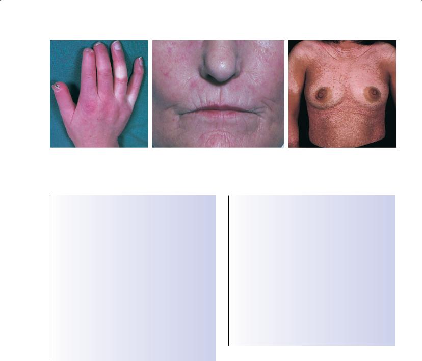
Figure 19.2 Characteristic features of limited cutaneous scleroderma. (a) Puffy fingers, tight skin, Raynaud’s phenomenon, loss of distal digits and ulceration of tips of digits. (b) Microstomia and telangiectasia. (c) Hypopigmentation caused by diffuse cutaneous scleroderma.
Box 19.2 Characteristic findings and suggested treatment for limited cutaneous scleroderma
Early stage (≤5 years after onset)
•Constitutional symptoms—fatigue common
•Skin thickening—no or minimal progression
•Organs affected—Raynaud’s phenomenon, ulcers of digital tips, oesophageal symptoms
•Treatment—vascular treatment (oral or intravenous) with or without digital sympathectomy, removal of calcinosis, treat oesophageal problems
Late stage (>10 years after onset)
•Constitutional symptoms—fatigue common and aggravated by effects of vasculopathy and gut disease
•Skin thickening—stable or slow progression
•Organs affected—Raynaud’s phenomenon, ulcers of digital tips, calcinosis, oesophageal stricture, small bowel malabsorption, pulmonary arterial hypertension, lung fibrosis
•Treatment—vascular treatment (oral or intravenous) with or without digital sympathectomy, removal of calcinosis, treat oesophageal and midgut problems
Box 19.3 Characteristic findings and suggested treatment for diffuse cutaneous scleroderma
Early stage (≤5 years after onset)
•Constitutional symptoms—fatigue, weight loss, pruritis
•Skin thickening—rapid progression with peak involvement by 2 years typical
•Organs affected—risk of renal, cardiac, pulmonary fibrosis, gastrointestinal, articular and muscular damage
•Treatment—vascular therapy, physiotherapy and occupational therapy as appropriate; immunosuppression for lung fibrosis and severe or progressive skin involvement; low-dose corticosteroids
Late stage (>5 years after onset)
•Constitutional symptoms—generally diminished
•Skin thickening—stable or regression
•Organs affected—musculoskeletal deformities, progression of existing visceral diseases but reduced risk of new complications
•Treatment—treat complications, gradual withdrawal of immunosuppression
around 18–30 months. Thereafter skin involvement tends to stabilize or improve. Despite stabilization or improvement in skin sclerosis, internal organ complications may develop at a later stage, and so long-term follow-up is mandatory. The characteristics of patients with each subset at different times in their disease are summarized in Boxes 19.2 and 19.3. Raynaud’s generally develops concurrently with the skin disease or shortly afterwards. The limited cutaneous subset of SSc (Figure 19.2) accounts for around 60% of cases in most North American or European series. Skin involvement is much less extensive and may be confined to the fingers (sclerodactyly), face or neck. Raynaud’s phenomenon is very prominent and may precede development of SSc by several years. The designation “CREST syndrome” is popular in the USA, referring to a subgroup of lcSSc in whom calcinosis, Raynaud’s
phenomenon, oesophageal dysmotility, sclerodactyly and telangiectasia occur. It is probably better not to distinguish such cases, as these manifestations are not universal and under-emphasize the life-threatening complications that develop in a significant proportion of lcSSc patients. These include pulmonary arterial hypertension, severe midgut disease and interstitial pulmonary fibrosis. Other SSc cases include overlap syndromes with features of polyarthritis, myositis or systemic lupus erythematosus, and the small group of SSc sine scleroderma who have major visceral involvement, Raynaud’s phenomenon and a hallmark autoantibody— typically anti-topoisomerase 1. The term “MCTD” is probably best avoided, as most of the patients with this designation evolve into a defined overlap syndrome, often with prominent features of SSc or lupus.
126 ABC of Rheumatology
(a) |
(b) |
(c) |
(d) |
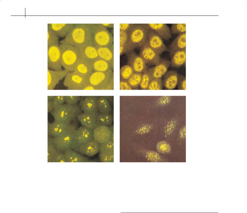
Figure 19.3 Common immunofluorescent patterns seen on testing for antinuclear antibodies. (a) Homogeneous—typical of antibodies to DNA, with or without histones. (b) Speckled—typical of antibodies to Ro, La, Sm and ribonucleotide protein. (c) Nucleolar—typical of scleroderma. (d) Centromere—mainly found with limited cutaneous scleroderma
Autoantibody profiles
The major hallmark autoantibodies associated with SSc are mutually exclusive (Figure 19.3). Thus, if a patient has anti-centromere antibodies they will almost never have anti-topoisomerase 1 or another reactivity associated with SSc. This appears to reflect the immunogenetic background of these individuals and may explain the clinical differences between patients with hallmark reactivity. The SSc-associated patterns of autoantibody reactivity are summarized in Table 19.2.
Risk stratification in SSc
The clinical heterogeneity of SSc and differences in natural history between the two major subsets and the life-threatening nature of some of the SSc-associated complications have led to attempts to risk-stratify patients at diagnosis and initial assessment. Abnormalities reflecting systemic inflammation (elevated erythrocyte sedimentation rate), pulmonary disease (impaired diffusion capacity, DLCO) or renal involvement (proteinuria) identifies patients with a poor 5-year survival. In addition, the clinical association of antibody profiles allows patients at increased risk of
Table 19.2 Autoimmune serology in SSc
Antibody |
Prevalence |
Comments |
|
|
|
ACA |
60% lcSSc |
Associated with typical |
|
|
CREST |
Scl-70 |
40% dcSSc, |
Predictive of interstitial lung |
|
15% lcSSc |
involvement, especially in |
|
|
lSSc |
RNApol |
20% SSc |
Anti-RNApol I or III associated |
|
|
with diffuse subset and renal |
|
|
disease |
U1-RNP |
10% SSc |
Associated with overlap features |
U3-RNP |
5% SSc |
Poor outcome and isolated PHT |
|
|
in dcSSc |
PM-Scl |
3% SSc |
Myositis overlap |
Th/To |
5% SSc |
Lung fibrosis in lcSSc |
anti-M2 |
5–10% SSc |
Especially in lcSSc with |
|
|
PBC |
|
|
|
See also chapters 18 and 20