Материал: ABC-of-Rheumatology-ABC-Series-

SLE and Lupus-like Syndromes |
117 |
|
|
|
|
ing accelerated hypertension and renal shutdown. Renal biopsy is helpful for assessing the severity, nature, extent and reversibility of the involvement and is an important guide to treatment and prognosis. For example, those with mesangial nephritis (class I) rarely progress to renal failure, in contrast to those with diffuse proliferative glomerulonephritis (class IV), who are at risk for end-stage renal disease.
Nervous system manifestations
SLE may affect the central and peripheral nervous systems. Definitions for these manifestations have been proposed by a consensus group (American College of Rheumatology, 1999) (Boxes 18.5 and 18.6). The most common manifestations are headache, seizures, aseptic meningitis and cerebrovascular accidents. Antiphospholipid antibodies (including anticardiolipin antibodies) have been implicated in cerebrovascular accidents and chorea. It often is hard to determine whether the depression and headaches are due to lupus itself; in many cases, they are related to psychosocial issues. Other possible causes such as sepsis, drugs, uraemia, severe hypertension and other metabolic causes must be sought and treated. Steroids are often blamed for inducing psychosis, but if any doubt exists, patients should be given more, not less, steroid while under medical supervision, particularly if active lupus is evident in other systems.
Box 18.5 Central nervous system manifestations of systemic lupus erythematosus
•Aseptic meningitis
•Cerebrovascular disease
•Demyelinating syndrome
•Headache (including migraine and benign intracranial hypertension)
•Movement disorders (including chorea)
•Myelopathy
•Seizure disorders
•Acute confusional state
•Anxiety disorder
•Cognitive dysfunction
•Mood disorder
•Psychosis
Box 18.6 Peripheral nervous system manifestations of systemic lupus erythematosus
•Acute inflammatory demyelinating polyradiculoneuropathy (Guillain–Barré syndrome)
•Autonomic disorder
•Mononeuropathy (single or multiplex)
•Myasthenia gravis
•Neuropathy, cranial
•Plexopathy
•Polyneuropathy
Box 18.7 Risk of atherosclerosis in lupus
Survival and disease control have improved in patients with SLE, but complications of premature vascular disease are recognized increasingly: the relative risk for myocardial infarction in women aged 35–44 years with SLE is 52.3 times the risk for women without lupus
Pulmonary and cardiovascular manifestations
Pleurisy, often without physical signs, is common in SLE. Less common manifestations are lupus pneumonitis, pulmonary haemorrhage, pulmonary embolism and pulmonary hypertension. Pulmonary haemorrhage can be sudden and acute and has high mortality. Pulmonary hypertension is associated with a poor prognosis, especially in pregnancy. Pericarditis is common but often asymptomatic. Other cardiac manifestations are myocarditis, endocarditis and rarely pericardial tamponade. Coronary artery disease is occasionally caused by vasculitis, but more often results from premature atherosclerosis (Box 18.7).
Gastrointestinal manifestations
Abdominal pain, nausea, vomiting and diarrhoea occur in up to 50% of SLE patients at some stage of the disease. Although the presentation of greatest importance is mesenteric vasculitis, in which the patient presents with an acute abdomen and is at high risk of death, there have been recent reports of patients with subacute abdominal pain or aseptic peritonitis. This is usually associated with other serological signs of active disease and generally improves with steroid therapy. Other abdominal manifestations include subacute bowel obstruction, hepatitis, sclerosing cholangitis, protein-losing enteropathy, pancreatitis and ascites. Exclusion or treatment of infection is essential in patients with these conditions.
Pregnancy and SLE
No evidence suggests that SLE reduces fertility, but active disease and the presence of antiphospholipid antibody syndrome (see below) may increase the risk of intrauterine growth retardation,
premature delivery, miscarriages and stillbirth (Gayed and Gordon, 2007). Doses of prednisolone >I0mg/day predispose to pre-
eclampsia, isolated hypertension in pregnancy, premature rupture of membranes and maternal infection. No evidence shows that
prednisolone crosses the placenta and causes fetal abnormalities in humans. Increasing evidence shows that azathioprine (<2mg/kg/
day) and hydroxychloroquine (200 mg daily) can be continued in pregnancy (Ostensen et al., 2006). If patients are on an angiotensinconverting enzyme (ACE) inhibitor or mycophenolate mofetil for renal disease, then medications must be discontinued due to their association with congenital malformations in exposed fetuses (Ostensen et al., 2006). Other anti-hypertensive medications such as methyldopa, labetalol and nifedipine are the most widely used to control blood pressure, and steroids and azathioprine can be added for lupus manifestations needing ongoing treatment.

118 ABC of Rheumatology
Box 18.8 Management of pregnancy
Pregnant women with lupus need close monitoring for optimal fetal and maternal outcome and are best managed in specialist units
Box 18.9 Management of congenital heart block
Most babies born with congenital heart block need a pacemaker during the first year of life
Box 18.10 Sjögren’s syndrome
Sjögren’s syndrome is often misdiagnosed as rheumatoid arthritis or systemic lupus erythematosus
ist advice, as they are at risk of systemic complications. Hydroxychloroquine and pilocarpine with other local symptomatic measures, such as artificial tears, are used to treat the condition.
Overlap syndromes and other lupus-like conditions
Up to 25% of patients with connective tissue disorders do not fit into classical descriptions and present with overlapping clinical features. Some may evolve into well-defined connective tissue disorders, while others have manifestations of more than one definite connective tissue disorder—e.g. systemic sclerosis combined with SLE and inflammatory myositis (see Chapter 21). Raynaud’s phenomenon is often present and may occur in isolation as the first manifestation of a connective tissue disorder. Patients with mild undifferentiated connective tissue disorders may have inflammatory arthritis, oedema of hands and acrosclerosis. Generally prognosis is good as long as patients do not develop pulmonary hypertension.
Pulmonary hypertension is associated with a 50% risk of mortality, particularly in the first 72 hours after delivery. This is usually a contraindication to planned pregnancy and needs specialist multidisciplinary care if diagnosed in pregnancy (Gayed and Gordon, 2007) (Box 18.8).
Neonatal lupus syndrome
This is a syndrome that occurs in about 10% of babies born to mothers with anti-Ro or anti-La antibodies. The most common manifestation is a rash induced by ultraviolet light a few days after birth. It resolves spontaneously if the babies are removed from sunlight or ultraviolet light. A more serious, but much rarer, manifestation of this syndrome is congenital heart block (CHB). This is usually detected in utero about 16–28 weeks into the pregnancy and may need treatment before or after delivery (Box 18.9).
Sjögren’s syndrome
This clinical syndrome is characterized by sicca symptoms: dry eyes and dry mouth due to failure of salivary and mucosal glands, often preceded by salivary gland swelling, and is associated with autoantibody formation as described below. It may occur as a secondary disorder in patients with SLE or other conditions, including rheumatoid arthritis, systemic sclerosis and primary biliary cirrhosis, or as a primary disorder with features that resemble a mild form of SLE (mild symmetrical arthritis, photosensitivity, fatigue and diffuse alopecia) (Box 18.10). The primary syndrome is associated with hypergammaglobulinaemia with very high total immunoglobulin G levels and definitely positive antinuclear antibody, rheumatoid factor, and anti-Ro and anti-La antibody tests. Patients with these immunological abnormalities may benefit from special-
Polymyositis and dermatomyositis
Proximal muscle weakness, elevated muscle enzymes, myopathic changes on electromyography and inflammatory changes on muscle biopsy are diagnostic criteria for polymyositis. The presence of a characteristic rash in the presence of the above features defines dermatomyositis. These diagnoses are made by fulfilling these criteria in combination and excluding other potential aetiologies for these test abnormalities.
Antiphospholipid syndrome
Antiphospholipid syndrome (Box 18.11) is an important cause of recurrent arterial and venous thrombosis and miscarriages that are associated with antiphospholipid antibodies (Box 18.12).
Thrombosis
The most common presentation of antiphospholipid syndrome is venous thrombosis in the arms or legs, which is often recurrent, multiple and bilateral, with a propensity for pulmonary embolism. Arterial thrombosis is less common but most frequently manifested by features of ischaemia or infarction. The severity of presentation depends on the acuteness and extent of the occlusion. The brain is the most common site, where thrombosis presents as stroke and transient ischaemic attacks. Other sites for arterial occlusion are the coronary arteries, and subclavian, renal, retinal and pedal arteries.
Obstetric syndromes
Recurrent pregnancy losses in the second or third trimester are typical (Box 18.11). Patients should be monitored for intrauterine growth restriction due to placental insufficiency and preeclampsia in a specialist unit. Planned early delivery is often required. (See below for treatment during pregnancy in the setting of antiphospholipid antibody syndrome.)
SLE and Lupus-like Syndromes |
119 |
|
|
|
|
Box 18.11 Criteria for classification of antiphospholipid syndrome
Clinical features
•Thrombosis
Confirmed episode of arterial and/or venous thrombosis in any organ or tissue
•Morbidity in Pregnancy
Fetal death beyond 10 weeks’ gestation with confirmed normal fetal morphology
Three or more spontaneous abortions before 10 weeks’ gestation in the absence of other maternal causes
More than one premature birth due to presence of severe placental insufficiency, pre-eclampsia or eclampsia before 34 weeks’ gestation
Laboratory criteria
•Immunoglobulin G and/or immunoglobulin M anticardiolipin antibodies in medium-to-high titre on at least two different
occasions more than 12 weeks apart (using a standard enzymelinked immunosorbent assay for β2-glycoprotein-I-dependent anticardiolipin antibodies) (Miyakis et al., 2006)
•Lupus anticoagulant in plasma on two separate occasions at least 12 weeks apart
Antiphospholipid syndrome definitely is present if at least one of the clinical features and one of the laboratory criteria are met
Box 18.12 Tests for antiphospholipid antibodies
•Anticardiolipin antibodies
•Antibodies against co-factors associated with anionic phospholipids, for example, β2-glycoprotein
•Lupus anticoagulant
•Biological false-positive serological tests for syphilis
Other manifestations
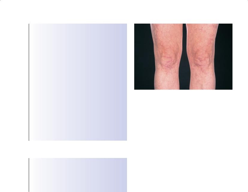
Other prominent features include thrombocytopenia (up to 50% of patients), haemolytic anaemia, livedo reticularis (Figure 18.4), chronic ulcers, typically near the medial malleolus, and cutaneous vasculitis.
Catastrophic antiphospholipid syndrome
This is an acute and devastating syndrome characterized by multiple simultaneous vascular occlusions throughout the body, which are often fatal. The kidney is affected most often, followed by the lungs, central nervous system, heart and skin.
Outcome of SLE and antiphospholipid syndrome
Although survival has improved substantially over the last 50 years (90% of patients survive at least 5 years and over 80% at least 10
Figure 18.4 Livedo reticularis
years), awareness is increasing that these patients succumb to late complications of the disease or its therapy. In particular, hyperlipidaemia, hypertension, premature ischaemic heart disease, diabetes mellitus and osteoporotic fractures may develop. Compliance with medications, clinic visits and lifestyle modifications is essential to prevent or reduce the risk of these associated problems, which may be iatrogenic or disease-related in origin (Bertsias et al., 2008). The long-term prognosis of antiphospholipid syndrome is poor, with organ damage in about one-third and functional impairment in up to one-fifth of patients at the end of 10 years.
Investigations
Investigations in SLE
A full blood count with differential white count, urinalysis and serum creatinine should be done for diagnosis and monitoring of the activity of SLE. Creatinine clearance or other assessment of glomerular filtration rate is more reliable for detecting early impairment of renal function. Patients with proteinuria or haematuria, or both, on dipstick must have microscopy done to look for casts if infection, stones and menstrual blood loss have been excluded.
For diagnosis, antinuclear antibody and anti-extractable nuclear antigen tests (see Chapter 24) should be done. No value is gained by repeating these tests, unless a change in clinical features is noted. Anti-ribonucleoprotein is associated with mixed connective tissue disease. Anti-dsDNA antibodies are useful for predicting patients at risk of developing renal disease and for monitoring disease activity. Although levels usually rise before a disease flare, they may fall at the time of flare. Levels of C3 and C4 fall with disease activity because of complement consumption, particularly in patients with renal disease. Levels also relate to the rate of synthesis in the liver and may rise in infections and pregnancy. Measurement of complement degradation products (for example, C3d, C4d) is less widely available but is more reliable for monitoring disease activity, as these reflect complement consumption alone. In women who are planning pregnancy, it is important to check for anti-Ro and anti-La antibodies and for antiphospholipid antibodies.

120 ABC of Rheumatology
Box 18.13 Confirmation of antiphospholipid antibody positivity
Tests for antiphospholipid antibodies must be positive on two separate occasions, at least 12 weeks apart, before a confident diagnosis of antiphospholipid syndrome can be made
Table 18.3 Management of systemic lupus erythematosus
Risk factor advice |
Further management |
Avoid sun and other ultraviolet light Avoid infection
Avoid unplanned pregnancy
Use non-steroidal anti-inflammatory drugs with care
Use oral steroids with care
Monitor for active disease
Screen for hypertension
Screen for diabetes and lipids
Assess osteoporosis risk
Wear sun block
Treat bacterial infections early with antibiotics
Advise appropriate contraception Use other analgesics as needed
Consider local and intramuscular or intravenous steroids and cytotoxic agents
Urinalysis, full blood count, creatinine, anti-dsDNA antibodies, C3, C4
Treat with calcium-channel blockers or angiotensinconverting enzyme inhibitors
Advise on diet and give drugs if needed
Give postmenopausal women bisphosphonates
Investigations in antiphospholipid syndrome
Overall, 80–90% of patients with antiphospholipid syndrome are positive for antibodies to a complex of anticardiolipin antibodies and β2-glycoprotein I. Lupus anticoagulant is only found in 20% of patients with antiphospholipid syndrome but is associated with a high risk of thrombosis. Low levels of antiphospholipid antibodies of no clinical consequence may develop transiently after infections (Boxes 18.12 and 18.13).
Management
General measures
Patients must be educated about the nature of their disease and the need for therapy. Leaflets from patient support organizations and references to reliable internet websites are useful. More than just drug therapy is required (Table 18.3; Box 18.14). Patients with sun-induced rashes should use sunblock regularly for about 6 months over the summer. Other patients with SLE should be aware that sun exposure may precipitate a disease flare.
Box 18.14 Lifestyle measures in the management of lupus
Lifestyle should be adjusted to ensure adequate rest, appropriate exercise, and a well-balanced diet. Measures for reducing stress should be considered and career plans reviewed. Patients with Raynaud’s phenomenon should wear appropriate warm clothing, including hats and gloves.
Table 18.4 Steroid-sparing and cytotoxic drugs used in systemic lupus erythematosus
Drug |
Range |
|
|
Hydroxychloroquine |
≤6.5 |
Azathioprine |
1–2.5 |
Methotrexate |
7.5–25 mg per week |
Cyclosporin A |
1–2.5 |
Lefluonamide |
10–20 mg daily |
Cyclophosphamide |
Intravenous pulses or ≤2 mg/kg/day orally |
Mycophenolate mofetil |
1–3 g/day |
|
|
Infections should be avoided and treated promptly if appropriate, as they can precipitate flares. Similarly, contraceptive pills that contain oestrogen may exacerbate lupus disease or thrombosis and should be used with caution. In general, barrier methods or progesterone-only contraception are preferred. Pregnancy should be planned, as the outcome is better, with fewer complications in both mother and fetus, if the mother has inactive disease at the time of conception. Drug therapy should be reviewed before conception.
Overlap and lupus-like conditions are managed in much the same way as mild SLE (Table 18.3; Box 18.14). Dry eyes should be managed by the frequent use of artificial tears. Dry mouth is best managed by taking sips of plain water, sucking ice cubes, or eating sugar free sweets. Artificial saliva preparations are disappointing.
Drug therapy in SLE
Milder cases with intermittent rashes, arthritis and other mucocutaneous features can usually be treated with steroid creams, short courses of non-steroidal anti-inflammatory drugs (NSAIDs) and hydroxychloroquine (<6.5 mg/kg/day). These drugs are also widely used in overlap syndromes, with the exception of NSAIDs, which are contraindicated in patients with features of systemic sclerosis or renal disease. More severe cases of SLE usually require oral corticosteroids. Patients who need 10 mg/day of prednisolone or more despite hydroxychloroquine, or those who present with more severe manifestations (such as nephritis, gastrointestinal vasculits or central nervous system disease) that need higher initial doses of prednisolone (0.5–1 mg/kg/day) are likely to need azathioprine, methotrexate or cyclophosphamide as steroid-sparing immunosuppressive agents (Table 18.4; Box 18.15). Mycophenolate mofetil

SLE and Lupus-like Syndromes |
121 |
|
|
|
|
Box 18.15 Cyclophosphamide therapy in severe lupus
Cyclophosphamide is often given as intermittent intravenous “pulse therapy” and is used predominantly for proliferative glomerulonephritis and systemic vasculitis
Box 18.16 Management of Raynaud’s phenomenon
Raynaud’s phenomenon is best treated with calcium-channel blockers, local nitrate creams if mild to moderate, and intravenous prostacyclin infusions in severe cases. Angiotensin-converting enzyme inhibitors may be tried if calcium-channel blockers are not tolerated.
is a promising alternative drug for the treatment of severe lupus; it has been best studied for lupus nephritis as an alternative to cyclophosphamide, but it is not licensed for SLE yet. Cyclosporin A, tacrolimus and lefluonamide are sometimes used for patients intolerant or resistant to other immunosuppressive agents (Table 18.4). Steroids should always be reduced slowly.
In pregnancy, patients may be given prednisolone, hydroxychloroquine and/or azathioprine, as the advantages are now considered to outweigh the risks. During lactation, prednisolone and hydroxychloroquine are acceptable, and azathioprine rarely causes problems at low doses. Methotrexate, mycophenolate, lefluonamide and cyclophosphamide are contraindicated in pregnancy and while breastfeeding. Cyclosporin A has been used in pregnancy in patients who have undergone transplants, but is not usually recommended during lactation.
Meticulous screening and treatment of blood pressure, diabetes, hyperlipidaemia and osteoporosis are essential. In general, cal- cium-channel blockers, ACE inhibitors and angiotensin receptor blockers are the preferred anti-hypertensive agents, as they are helpful in the management of Raynaud’s phenomenon and renal disease, and because β-blockers aggravate Raynaud’s phenomenon (Box 18.16). Bisphosphonates are often required in postmenopausal women, but they should be used with great care in women who may want to become pregnant in the future. Bisphosphonates, statins, ACE inhibitors and angiotensin-receptor blockers should be stopped before a planned pregnancy. Calcium and vitamin D can be used in all age groups. Treatment with anticoagulation and anti-epileptic, antidepressant, or antipsychotic drugs should be considered early in the management of patients with neuropsychiatric disease.
Oestrogen-containing contraceptives and hormone replacement therapy should be used with care in women with stable mild/moderate lupus and should be avoided in women with antiphospholipid antibodies, especially those with a history of thrombosis or pregnancy loss. Progesterone-only contraception is acceptable but is associated with a theoretical increased osteoporotic risk. Intrauterine devices can be used in women in stable relationships with a low risk of infection.
Box 18.17 Goals of treatment in antiphospholipid syndrome
•Prophylaxis
•Treatment of acute thromboses
•Prevention of further thrombotic events
•Management of pregnancy in antiphospholipid syndrome
Therapy in antiphospholipid antibody syndrome
There is no evidence for prophylactic treatment of patients serologically positive for antiphospholipid antibody syndrome but without a history of thrombosis, although aspirin is often given in practice. Oestrogen-containing contraceptives and hormone replacement therapy are best avoided in patients with antiphospholipid syndrome and in patients with antiphospholipid antibodies without thrombosis or fetal loss. Treatment for thrombosis is usually initiated with intravenous or subcutaneous heparin and is soon changed to oral anticoagulation with warfarin. Most doctors recommend maintaining the international normalized ratio (INR) at 2–3 to prevent venous thrombosis and between 3 and 4 to prevent arterial thrombosis, as studies suggesting that INR in the range 2–3 was sufficient for patients after arterial thrombosis were flawed. Anticoagulation is usually life-long unless a contraindication, such as poorly controlled hypertension, is present (Box 18.17).
Pregnancy—A combination of low-molecular-weight heparin and low-dose aspirin is preferred. Pregnant women need close monitoring by the obstetrician, haematologist and rheumatologist, preferably in combined clinics at specialist units. Heparin dosage will depend on the clinical circumstances (Derksen et al., 2004).
Conclusion
SLE is more common than many people realize. The presentations are diverse, and it may take a few years to realize that a variety of symptoms and signs can all be attributed to SLE, Sjögren’s syndrome or an overlap syndrome. Antiphospholipid antibody syndrome should be sought actively in patients with a history of recurrent fetal loss or thrombosis, or both, because of the risk of future thrombotic complications. These diagnoses should not be made without appropriate clinical and serological features, as there are many social consequences of these diagnoses, such as implications for obtaining insurance and mortgages. With appropriate treatment, the outcome of these conditions is good, but the risk of late complications, particularly those of atherosclerosis, is important. In future, the management of patients with these conditions should seek to reduce these risks as well as control active disease.
References
American College of Rheumatology. American College of Rheumatology nomenclature and case definitions for neuropsychiatric lupus syndromes.
Arthritis and Rheumatism 1999; 42: 599–608.