Материал: ABC-of-Rheumatology-ABC-Series-

CHAPTER 17
Polymyalgia Rheumatica and Giant
Cell Arteritis
Eric L Matteson1 and Howard A Bird2
1Mayo Clinic, Rochester, USA
2Chapel Allerton Hospital, Leeds, UK
OVERVIEW
•Patients with polymyalgia rheumatica are over 50 years of age, and on average over 70 years old.
•Hallmark symptoms of the disease are shoulder and hip girdle pain with marked stiffness.
•Giant cell arteritis may be present in at least 30% of patients.
• Treatment is with glucocorticosteroids, initially 15–20 mg a day of prednisone equivalent. Treatment is often required for several years.
Polymyalgia rheumatica (PMR) is a clinical syndrome that affects older patients and comprises proximal muscle group stiffness, particularly in the shoulder, and systemic features such as fatigue and weight loss. It is associated with an increased erythrocyte sedimentation rate (ESR) and responds dramatically to relatively small doses of steroids.
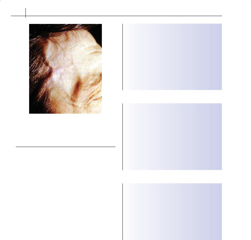
Giant cell arteritis (GCA) is a systemic vasculitis that affects largeand medium-sized arteries. Although it may involve any artery, it has a propensity to affect the branches of the external carotid artery, particularly the posterior ciliary arteries that supply the optic nerve and the superficial temporal artery; hence its alternative (often interchangeable) name “temporal arteritis” (Figure 17.1).
There are clinical and pathogenetic links between temporal arteritis, GCA and PMR, which has led to the concept that they are manifestations of a disease spectrum that affects the same disease population (Box 17.1). The two entities may occur in the same patient simultaneously, at different time points, or independently. PMR has been observed in 40–60% of cases of GCA, and 30–80% of patients with PMR have GCA.
Causes
The cause of GCA or PMR is likely to be polygenic, with both genetic and environmental factors contributing to disease susceptibility and severity.
ABC of Rheumatology, 4th edn. Edited by Ade Adebajo. ©2010 Blackwell Publishing Ltd. 9781405170680.
Environmental
Acute-onset prodromal events and synchronous variations in incidence of PMR and GCA suggest a possible environmental infectious trigger. Several studies have shown concurrence in the incidence of PMR and GCA with epidemics of Mycoplasma pneumoniae, Chlamydia pneumoniae, parvovirus B19, respiratory syncytial virus and adenovirus. Despite this, no definite causative infectious agent has been identified.
Genetics
Racial differences in incidence and familial aggregation suggest a common genetic susceptibility factor. PMR and GCA are linked with human leukocyte antigen DR4 (HLA-DR4). Patients who are positive for HLA-DR4 and those who are negative for HLA-DR4 do not present differently. A conserved sequence within the second hypervariable region located in the antigenbinding groove of the HLA-DR molecule has been identified. Differential expression of genes responsible for inflammatory cytokine expression are likely to account for the variable disease manifestations.
Clinical features
In the absence of a specific diagnostic test, apart from biopsy of the temporal artery when GCA is also present, the diagnosis of PMR is based on clinical features (Table 17.1) and made by exclusion
Box 17.1 Epidemiology of polymyalgia rheumatica and giant cell arteritis
•Giant cell arteritis is the most common of the vasculitides, and polymyalgia rheumatica is more common than giant cell arteritis
•Pre-eminently affect Northern European people; can occur in any ethnic group
•Rare under the age of 50 years
•Woman:man ratio = 3:1
•Annual incidence approximately 18 per 100,000 for giant cell
arteritis and 100 per 100,000 for polymyalgia rheumatica in people aged >50 years
•Possible cyclic pattern in incidence
•Siblings at increased risk
107
108 ABC of Rheumatology
Figure 17.1 Markedly dilated temporal arteries in a 74-year-old man with giant cell arteritis. The arteries are visibly thickened and inflamed; palpation of the vessel is painful. Courtesy of Dr Lester Mertz, Mayo Clinic, Rochester, MN, USA
Table 17.1 Clinical features
Polymyalgia rheumatica |
Giant cell arteritis |
|
|
Bilateral shoulder pain, stiffness |
Age >50 years |
Duration onset <2 weeks |
New headache |
Initial ESR >40 mm/hour |
Temporal artery abnormal |
Stiffness >1 hour |
to palpation |
Age >50 years, typically >65 years |
Elevated ESR |
Depression or weight loss, or both |
Abnormal findings on |
Bilateral upper arm tenderness |
temporal artery biopsy |
Probable PMR: 3 or more <3 with clinical |
|
abnormality of temporal artery |
|
Definite PMR: probable PMR responding to |
|
corticosteroids |
|
|
|
ESR = erythrocyte sedimentation rate; PMR = polymyalgia rheumatica
(Boxes 17.2 and 17.3). Several diagnostic criteria sets have been suggested.
In PMR, the onset is relatively rapid, over a matter of days to several weeks. The shoulder pain and stiffness are invariably bilateral and can be profound. Acute-phase reactants such as ESR and C-reactive protein (CRP) are usually (>95%) elevated (Box 17.4). Systemic features may include low-grade fever, fatigue, weight loss and depression. Sometimes, the muscles of the upper arms and thighs are tender on direct palpation, although muscle strength is usually unimpaired. Patients may have difficulty turning over in bed, particularly early in the morning. It may be hard to lift heavy objects, painful to walk up stairs, or tender to sit on the toilet. Patients comment on their “overwhelming illness”. Unusual upper-arm tenderness when blood pressure is taken should raise suspicion of the disease, especially in patients with other constitutional symptoms.
Box 17.2 Polymyalgia: rarer mimics
•Polyarteritis nodosa
•Parkinson’s disease
•Thyrotoxic myopathy and hypothyroidism
•Carcinomatous myopathy
•Systemic lupus erythematosus
•Calcium pyrophosphate deposition
•Multiple myeloma
•Paget’s disease
•Osteomalacia
•Polymyositis
•Subacute bacterial endocarditis
•Malignancy
Box 17.3 Polymyalgia rheumatica: differential diagnoses
•Polymyalgia
•Osteoarthritis
•Cervical spondylosis
•Frozen shoulder
•Rheumatoid arthritis
•Giant cell arteritis
•Cluster headache
•Non-inflammatory ophthalmic ischemia
•Systemic amyloidosis
•Other forms of vasculitis, including polyarteritis nodosa
•Occipital myalgia
•TMJ syndromes
Box 17.4 Markers of disease activity
Although there are numerous non-specific acute-phase reactants such as heptoglobin and cytokines such as interleukin-6 and vascular endothelial growth factor, as well as plasma viscosity, which may be elevated in polymyalgia rheumatica/giant cell arteritis, only two have routine clinical utility:
•Erythrocyte sedimentation rate
•C-reactive protein
•Other common laboratory abnormalities include a non-specific normochromic, normocytic anaemia and elevation of alkaline phosphatase (bone and/or liver fraction)
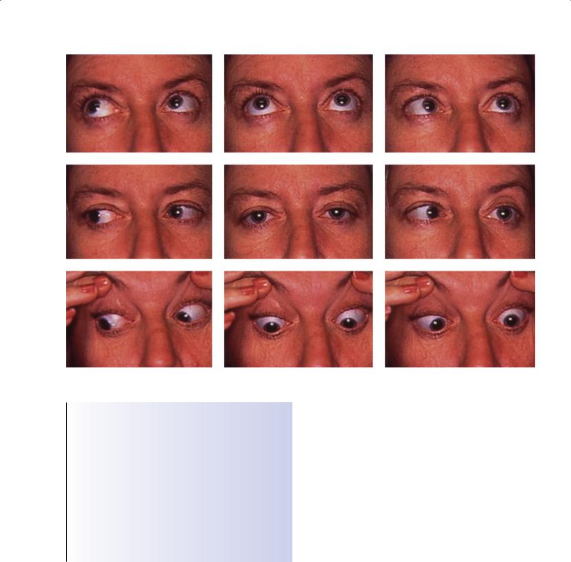
GCA usually may be viewed as inflammation of the aorta and its major branches. Its clinical features are related to the affected arteries. The scalp is tender to the touch, and it may even hurt to wear spectacles. Jaw claudication may occur while chewing. Clinical signs (Table 17.1) vary according to the duration of the disease. In the early stages, the pulse is full and bounding, and the arteries tender. Later, fibrosis and repair may predominate, the artery may have a nodular indurated feel to it, and the pulse is almost absent. Diplopia, partial or complete loss of vision, and cranial nerve palsy
PMR and GCA |
109 |
|
|
|
|
Figure 17.2 Temporary or permanent nerve palsy can occur in association with cranial arteritis. In this case, the sixth nerve is clearly involved
Box 17.5 Biopsy for giant cell arteritis
•Biopsy is most useful just before or within 24 hours of treatment initiation with steroids, but treatment should not be delayed for the sake of obtaining a biopsy
•Skip lesions occur, so a negative result does not exclude giant cell arteritis
•A positive result may resolve later doubt about diagnosis, particularly if the response to treatment is not rapid and classical
•It may not be possible to biopsy all patients; the decision depends on local resources
•One week after starting steroid treatment, the chance of
obtaining positive biopsy falls to 10%, although the biopsy may still reveal evidence of inflammation >1 year after initiation of
treatment
(Figure 17.2) may all occur if the condition remains untreated. Late complications of large-vessel involvement, including aortic aneurysm and stenosis, may complicate the disease course. Patients should be followed long term for aortic disease with computed tomography and aortic magnetic resonance imaging (MRI) and complemented by ultrasonography of the aortic root and abdominal aorta by clinical and imaging assessment, as aneurysmal rupture is a cause of premature mortality in these patients. The value of biopsy for GCA is discussed in Box 17.5.
Histopathology
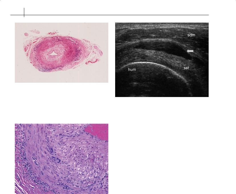
Giant cell arteritis—The inflammatory features of GCA are typically described as illustrating the “skip” phenomenon due to the patchy or segmental involvement of the arteries. GCA is principally a disease driven by T-cells that is limited to vessels with an internal elastic component. Histologically, the lesions are characterized by a mainly lymphocytic and macrophage infiltrate with the presence of giant and epithelioid cells (Figure 17.3). The CD3+ and T-cell population comprises CD4+ or CD8+ subsets, of which CD4+ T-cells predominate. The initial immunological event—probably the induction of CD4+ T-cell proliferation by an unknown antigen—occurs in the outer vessel layer: the adventitia. These CD4 cells produce interferon-©, which attracts macrophages to the arterial wall, where they fuse to form multinucleated giant cells in the intima-media junction (Figure 17.4). The giant cells produce express adhesion molecules, nitric oxide and collagenases to result in, for example, tissue injury and in situ thrombosis.
Polymyalgia rheumatica—The histopathological features of PMR are defined less clearly than for GCA. Biopsy of synovium, especially of shoulder-joint structures, have confirmed synovitis in about one-third of patients, which is nonerosive and self-limiting arthritis.
110 ABC of Rheumatology
Figure 17.3 Histology of temporal arteritis. The elastic tissue appears black, while various types of collagen stain yellow. Remnants of the internal elastic lamina are indicated by an arrow. An inflammatory infiltrate is found in the media and intimal fibrosis. Multinucleated giant cells and macrophages are attacking the elastic tissue and ingesting it. There is extensive intimal proliferation and fibrosis. Luminal narrowing has occurred almost completely. Involvement of other arteries may occur, including the ophthalmic artery, which results in loss of vision
Figure 17.5 Ultrasound examination of a patient with polymyalgia rheumatica and subdeltoid bursitis. Anterior transverse ultrasound image of the right shoulder with maximum internal rotation of the arm: the dark area (arrow) between the subdeltoid muscle and supraspinatus tendon represents subdeltoid bursitis. hum = humerus; sdm = subdeltoid muscle; sst = supraspinatus tendon. Courtesy of Dr Wolfgang Schmidt, Humboldt University, Berlin, Germany
Figure 17.4 Photomicrographs of active arteritis in a temporal artery biopsy. High-power view showing proliferation of intimal fibroblasts and transmural inflammation with multinucleated giant cells present at the media-intima junction (Hematoxylin-Eosin, 200×)
Inflammation of the temporal artery may also be demonstrated even in patients without overt GCA. The characteristics of synovitis on biopsy confirm a predominance of CD4+ T-cells and macrophages. Similar to features described in GCA, the vascular infiltrate reveals CD4+ interferon-©-positive cells in the adventitia; macrophages that produce interleukin-1®, interleukin-6 (IL-6), endothelial cell adhesion molecules with matrix metalloproteinases, and inducible nitric oxide are seen in the media intima.
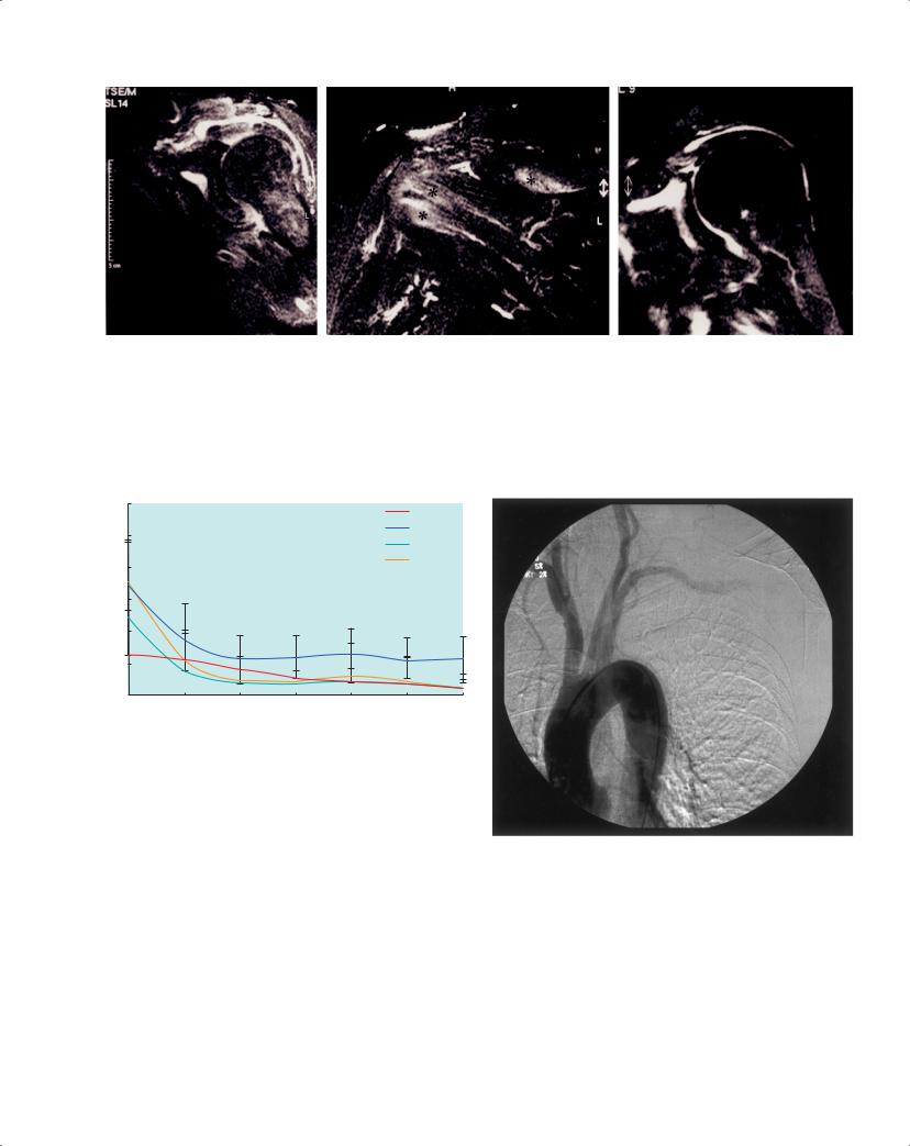
Increasingly used in clinical evaluation, ultrasonography of the shoulders (Figure 17.5) and hips may reveal synovitis of the joints or bursa. MRI studies also give evidence of an inflammatory process affecting distal articular or extra-articular (tenosynovial) structures, or both (Figure 17.6). Although skeletal muscle is not
considered to be a site of pathology, focal changes in muscle ultrastructure and mitochondria abnormalities have been noted, but their significance remains unclear. Muscle enzymes and biopsies are normal.
Investigations
ESR and/or CRP are the most accepted and easily available markers of active inflammation in PMR (Figure 17.7). They are initially elevated in over 90% of patients, although PMR and even blindness from GCA can occur in the presence of a normal ESR. The ESR and CRP fall with effective treatment; the CRP falls faster. With treatment, the normocytic normochromic anaemia corrects, and the slight increase in hepatic alkaline phosphatase sometimes noted in active disease is reduced.
In parallel, to these basic laboratory studies, additional investigations should be arranged to exclude conditions that cause diagnostic confusion, including thyroid function studies and ageand symptom-appropriate malignancy screening. Additionally, an evaluation should be performed in all patients to screen and follow common conditions of elderly patients, which may be induced or exacerbated by protracted corticosteroid use, including diabetes, osteoporosis and cardiovascular disease.
Vascular assessment with ultrasound, computed tomography/ MRI, or conventional angiography may be required to assess the activity and extent of vascular involvement (Figure 17.8). Ultrasound and MRI may reveal a characteristic “halo” around inflamed vessels, even of the caliber of the temporal arteries. Positron emission tomography may occasionally be useful in defining disease activity, but remains experimental.
The basic investigations described above are summarized in Box 17.6.
PMR and GCA |
111 |
|
|
|
|
Figure 17.6 MRI of shoulder in polymyalgia rheumatica (T2 fat suppressed corona). Oblique MRI sequences shows modest synovial inflammation with substantial fluid in the subacromial bursa associated with diffuse capsular edema extending into the adjacent tendons and muscle bellies. Courtesy of Dr D. McGonagle and Dr H. Marzo-Ortega, Academic Unit of Musculoskeletal Disease, Leeds General Infirmary, Leeds, UK
120
Corticoid
ESR
100
CRP
VAS pain
80
60
40
20
0
1 |
2 |
3 |
4 |
5 |
6 |
7 |
Weeks
Figure 17.7 Improvement in erythrocyte sedimentation rate, C-reactive protein, and the patient’s perception of pain (measured on a visual analogue scale) in response to corticosteroid therapy in a group of 76 patients with polymyalgia rheumatica. Courtesy of Dr Burkhard Leeb, Lower Austria Centre for Rheumatology, Stockerau, Austria
Figure 17.8 Arterial thrombosis complicating polymyalgia rheumatica, supporting a generalized vasculitic aetiology of this condition. Courtesy of Dr C. Pease, Academic Unit of Musculoskeletal Disease, Leeds General Infirmary, Leeds, UK