Материал: ABC-of-Rheumatology-ABC-Series-

Juvenile Idiopathic Arthritis |
97 |
|
|
|
|
otrexate) to control features of systemic arthritis. It is initially well tolerated but only anecdotally successful. It has also been combined with methotrexate to treat uveitis. Side effects include gingival hyperplasia and hirsuitism. Over time, cyclosporin A contributes to hypertension and progressive renal disease. It is still used routinely to treat macrophage activation syndrome associated with systemic arthritis either solo or in combination with high-dose methylprednisolone.
Cyclophosphamide—Borrowing from oncology, high doses of chemotherapy are sometimes advocated for patients unresponsive to all other available therapies, particularly children with severe recalcitrant systemic arthritis whose quality of life is poor and for whom even the newest biologic therapies are not working.
Autologous stem-cell transplantation
A number of children with truly recalcitrant systemic arthritis have been successfully transplanted in Europe, and the procedure is being investigated in the USA for JIA unresponsive to all other therapies. It is a high-risk procedure but seems to offer a reasonable chance of inducing disease remission.
Natural history
JIA is a diverse group of conditions, each unique but all associated with persistent arthritis. The spectrum of JIA varies from: mild to severe; smouldering to rapidly progressive; uniphasic and polyphasic to chronic and continuous; affecting one to affecting most joints; associated with no extra-articular symptoms to such severe extra-articular manifestations as to overshadow the presence of arthritis. Accordingly, few children may remit spontaneously (predictive factors for this are unknown), while another small fraction will have devastating disease, recalcitrant to all, including experimental, treatments. For the remaining majority of children with JIA, however, long-term follow-up studies highlight a much poorer prognosis than previously believed (Foster et al., 2003). These studies show a profound impact on later productivity and on quality of life of adults diagnosed at a time when biologic therapies had not yet been found. It follows then that early diagnosis and rapid referral to an experienced paediatric rheumatology team would be associated with improved outcome and that the longer duration of inflammation, the higher the impact on quality of life, in particular on independence in activities of daily living.
Studies of large numbers of children with JIA followed longitudinally show that as many as 30% continue to have active arthritis
into their adult years. These studies include children with persistent oligoarthritis whose natural history of illness has been well described and tends to be limited to an average of 2 years of monoarthritis. That leaves children with polyarthritis and systemic arthritis, in addition to 20% with extended oligoarthritis, comprising the group who continues to have disease into adulthood. Today, many of these young adults are left with the chronic sequelae of short stature, restricted joint movement, asymmetrical growth and extraarticular abnormalities. An appreciation of the true natural history of JIA and the availability of successful treatments have imparted great urgency to prompt referral, initiation of aggressive treatment regimens, improved access to clinical trials and renewed hope for children with arthritis. Over the next decade, new treatments will be developed and tested in children, while concomitant translational research, including studies in pharmacogenetics, will all result in custom-designed individualized and uniquely targeted therapies.
References
Foster HE, Marshall N, Myers A, Dunkley P, Griffiths ID. Outcome of adults with juvenile idiopathic arthritis; quality of life study. Arthritis and Rheumatism 2003; 48: 767–775.
Lovell DJ, Giannini EH, Reiff A et al. for the Pediatric Rheumatology Collaborative Study Group. Long-term efficacy and safety of etanercept in children with polyarticular course juvenile rheumatoid arthritis. Arthritis and Rheumatism 2003; 48: 218–226.
Silverman E, Mouy R, Spiegel L et al. Leflunomide or methotrexate for juvenile rheumatoid arthritis. New England Journal of Medicine 2005; 352: 1655–1666.
Further reading
Brooks CD. Sulfasalazine for the management of juvenile rheumatoid arthritis. Journal of Rheumatology 2001; 28: 845–853.
Jung JH, Jun JB, Yoo DH, Kim TH, Jung SS, Lee IH et al. High toxicity of sulfasalazine in adult-onset Still’s disease. Clinical and Experimental Rheumatology 2000; 18: 245–248.
Ruperto N, Lovell DJ, Cuttica R et al. for the Pediatric Rheumatology International Trials Organisation and the Pediatric Collaborative Study Group. A randomised placebo controlled trial of infliximab plus methotrexate for the treatment of polyarticular-course juvenile rheumatoid arthritis. Arthritis and Rheumatism 2007; 56: 2815–2816.
Szer IS, Kimura Y, Malleson PN, Southwood TR, eds. Arthritis in Children and Adolescents: juvenile idiopathic arthritis. Oxford University Press, Oxford, 2006.

CHAPTER 16
Musculoskeletal Disorders in Children
and Adolescents
Helen Foster1 and Lori Tucker2
1Newcastle University, Newcastle upon Tyne, UK
2British Columbia’s Children’s Hospital, Vancouver, Canada
OVERVIEW
•Musculoskelelal complaints in children are common, often benign and self-limiting, but can be presenting features of significant, severe and potentially life-threatening conditions.
•Making a diagnosis rests on competent clinical skills, knowledge of normal variants, knowledge of common clinical scenarios, “red flags” to suggest severe conditions and judicious use and interpretation of investigations.
•Common clinical scenarios include the limping child, “growing pains”, back pain and knee pain.
•Knowledge of “red flags” to suggest infection, malignancy, multi-system disease and inflammatory joint disorders are important.
•The management of musculoskeletal conditions involves a multidisciplinary approach.
•Many chronic conditions that begin in childhood continue into adult life, and the process of transitional care to adult services starts in early adolescence.
Introduction
Musculoskeletal (MSK) presentations in childhood are common, with a spectrum of causes (Box 16.1), the majority of which are benign and self-limiting. It must be remembered, however, that severe, potentially life-threatening conditions such as malignancy, sepsis, vasculitis and non-accidental injury may also present with MSK complaints. Furthermore, MSK features are common in association with chronic conditions other than rheumatic disorders, such as inflammatory bowel disease and cystic fibrosis. Diagnosis relies on competent MSK clinical skills, with the minimum of a screening MSK examination (Foster et al., 2006), appropriate knowledge of normal variants (Box 16.2), “red flags” to raise suspicion of malignancy or sepsis and clinical scenarios at different
ABC of Rheumatology, 4th edn. Edited by Ade Adebajo. ©2010 Blackwell Publishing Ltd. 9781405170680.
Box 16.1 Differential diagnosis of musculoskeletal pain
Life-threatening conditions
•Malignancy (leukaemia, lymphoma, bone tumour)
•Sepsis (septic arthritis, osteomyelitis)
•Non-accidental injury
Joint pain with no swelling
•Hypermobility syndromes
•Idiopathic pain syndromes (reflex sympathetic dystrophy, fibromyalgia)
•Orthopaedic syndromes (e.g. Osgood–Schlatter disease, Perthes disease)
•Metabolic (e.g. hypothyroidism, lysosomal storage diseases)
Joint pain with swelling
•Trauma
•Infection
Septic arthritis and osteomyelitis (viral, bacterial, mycobacterial)
Reactive arthritis (post-enteric, sexually acquired)
Infection related (rheumatic fever, post-vaccination)
•Juvenile idiopathic arthritis
•Arthritis related Inflammatory bowel disease
•Connective tissue diseases (SLE, scleroderma, dermatomyositis, vasculitis)
•Sarcoidosis
•Metabolic (e.g. osteomalacia, cystic fibrosis)
•Haematological (e.g. haemophilia, haemoglobinopathy)
•Tumour (benign and malignant)
•Chromosomal (e.g. Downs related arthritis)
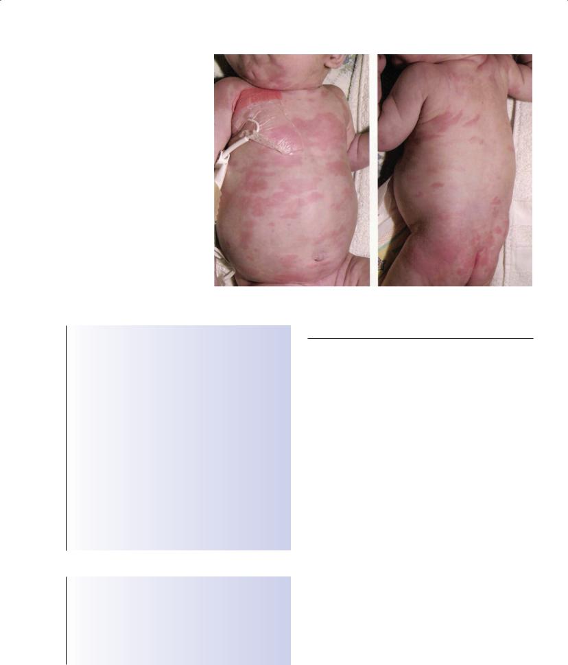
•Auto-inflammatory syndromes e.g. CINCA, (Figure 16.1) periodic syndromes, CRMO)
•Developmental/congenital (e.g. spondylo-epiphyseal dysplasia)
ages (Box 16.3). The approach to MSK assessment in children is different to that of adults; as young children may have difficulty in localizing or describing symptoms, the history is often given by the parent/carer, and complaints may be non-specific, such as “my child is limping”. Clinical assessment usually distinguishes between mechanical and inflammatory problems and an approach to assess-
98
MSK Disorders in Children and Adolescents |
99 |
|
|
|
|
Figure 16.1 Chronic infantile neurological cutaneous arthritis (CINCA) syndrome: widespread rash
Box 16.2 Normal variants in gait patterns and stance
•Intoeing can be due to:
Hip—persistent femoral anteversion—commonly between ages of 3–8 years
Lower leg—(internal tibial torsion)—commonly from onset of walking to 3 years
Feet—metatarsus adductus—most resolve by the age of 6 years
•Bow legs (varus)—birth to early toddler—most resolve by 3 years
•Knock knees (valgus)—most resolve by age of 7 years
•Flat feet—most resolve by 6 years, and normal arches are evident on tiptoeing
•Crooked toes—most resolve with weight bearing
Indicators to cause concern
•Persistent changes that fail to resolve by the expected age
•Progressive changes
•Lack of symmetry
•Associated pain or functional disability
•Systemic upset
•Dysmorphic features/short stature
Table 16.1 A strategy for characterizing musculoskeletal pain in children
Localized pain |
Diffuse pain |
|||
|
|
|
|
|
|
|
|
|
|
“Well” child |
“Unwell”** |
“Well” Child |
“Unwell” Child |
|
|
child |
|
|
|
|
|
|
|
|
Strains and sprains |
Septic arthritis |
Hypermobility |
Leukaemia |
|
Bone tumours |
Osteomyelitis |
Diffuse idiopathic |
Neuroblastoma |
|
JIA (oligoarticular |
|
|
pain syndromes |
JIA (systemic and |
subtype) |
|
|
|
polyarticular |
Localized |
|
|
|
onset subtypes) |
idiopathic pain |
|
|
|
SLE |
syndromes |
|
|
|
Juvenile |
“Growing pains” |
|
|
|
dermatomyositis |
|
|
|
|
Vasculitis |
|
|
|
|
|
**associated with one or more “red flags”, such as fever, anorexia, weight loss, malaise and raised inflammatory markers
JIA = juvenile idiopathic arthritis; SLE = systemic lupus erythematosus This table is adapted from Malleson and Beauchamp, 2001
Box 16.3 “Red flags” to warrant concern in children presenting with musculoskeletal symptoms
•Systemic upset (fever, malaise, anorexia, weight loss or raised inflammatory markers)
•Bone pain and/or night pain
•Regression of motor milestones
•Functional disability
ment (Table 16.1) incorporates potential diagnoses according to whether pain is localized or diffuse, whether the child is “well” or not and the presence or absence of “red flags”. The presence of multi-system features broadens the differential to include connective tissue diseases.
The “limping child”
This is a common presentation, with a spectrum of age-related
100 ABC of Rheumatology
Table 16.2 Common/significant causes of limping according to age
Toddler/preschool
Infection (septic arthritis, osteomyelitis—hip, spine) Mechanical (trauma and non-accidental injury) Congenital/developmental problems (e.g. hip dysplasia, talipes) Neurological disease (e.g. cerebral palsy, hereditary syndromes) Inflammatory arthritis (JIA)
Malignant disease (e.g. leukaemia, neuroblastoma)
5–10 years
Mechanical (trauma, overuse injuries, sport injuries)
Reactive arthritis/transient synovitis—”irritable hip”
Perthes’ disease
Inflammatory arthritis (JIA)
Tarsal coalition
Idiopathic pain syndromes
Malignant disease
10–17 years
Mechanical (trauma, overuse injuries, sport injuries) Slipped capital femoral epiphysis
Inflammatory arthritis (JIA) Idiopathic pain syndromes Osteochondritis dissecans Tarsal coalition
Malignant disease (leukaemia, lymphoma, primary bone tumour)
JIA = juvenile idiopathic arthritis
Reproduced by kind permission of the Arthritis Research Campaign (www. arc.org.UK)
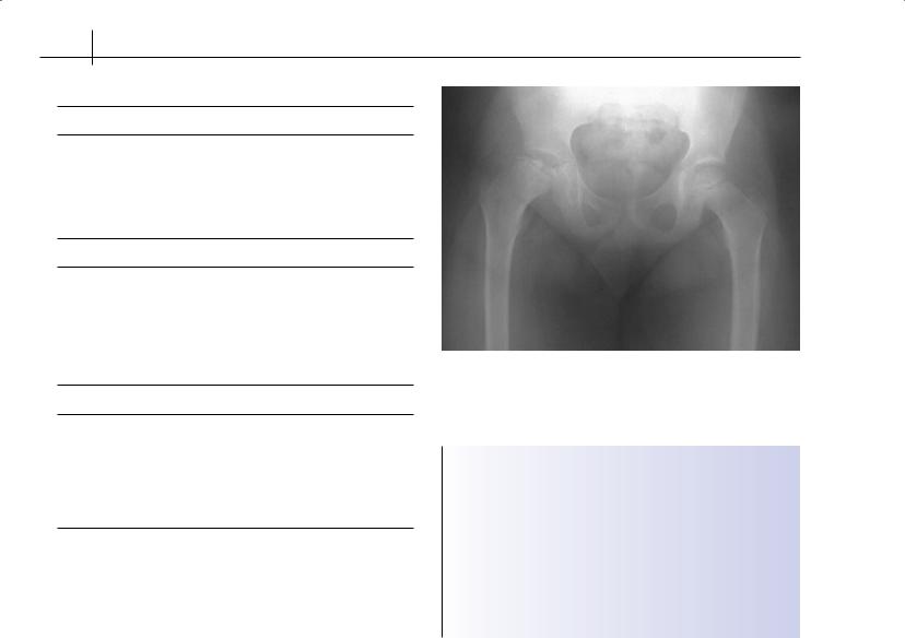
Figure 16.2 Perthes’ disease, showing avascular necrosis of the right hip
Box 16.4 The “rules” of growing pains
•Pains are never present at the start of the day after waking
•The child does not limp
•Physical activities are not limited by symptoms
•Pains are symmetrical in the lower limbs
•Physical examination is normal (evidence of joint hypermobility is common)
•The child is systemically well
•Gross motor milestones are normal
•Age range 3–11 years
causes (Table 16.2), and the site of the problem may be broad (from a foreign body in the sole of the foot to a tumour in the spine). Orthopaedic conditions at the hip are common and often present acutely, with a well (albeit limping) child, with possible diagnoses including slipped upper femoral epiphysis (usually the older, often overweight, child) and Perthes’ disease (Figure 16.2) (which may follow a transient synovitis or “irritable hip” in the younger child). The hip joint is unusual as a monoarthritis in juvenile idiopathic arthritis (JIA), and in isolation, sepsis (including mycobacterial infection) needs to be considered. The concept of referred pain from the hip or thigh, for example, must be sought in situations where the child has knee pain but there is no evidence of localized disease at the knee.
Back pain
Back pain is a common complaint, and is frequently mechanical with contributory factors such as poor posture, physical inactivity or abnormal loading (such as carrying heavy school bags on one shoulder). Certain sporting activities such as cricket, bowling or gymnastics pose increased risk of back pain, with possible consequences such as spondylolysis and spondylolisthesis. “Red flags” for referral for a child with back pain include a painful scoliosis,
neurological symptoms suggestive of nerve-root entrapment or cord compression and systemic findings to suggest malignancy or sepsis. Inflammatory back pain may be a late feature of enthes- itis-related arthritis (a subtype of JIA), often presenting in late adolescence and with a strong association with expression of HLA-B27.
Mechanical pain
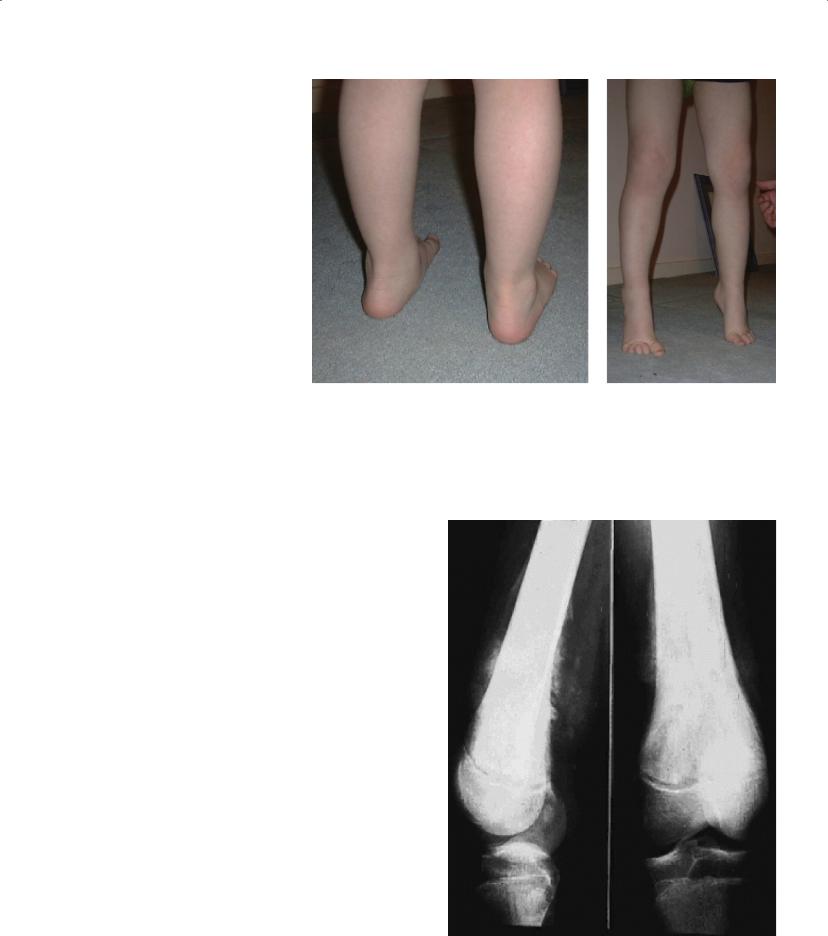
Osteochondritis of the knee (Osgood–Schlatter disease) is common, especially in adolescent boys who are physically active (particularly those who play football or basketball). Sever’s disease (osteochondritis of the calcaneum) may present with a painful heel. Flat feet (Figure 16.3) are common, and standing on tiptoe should create a normal medial longitudinal arch; inability to do so or painful fixed flat feet warrant further investigation to exclude tarsal coalition. High fixed arches, or pes cavus, may suggest neurological disease. Non-specific mechanical MSK pain in children is often labelled as “growing pains”. Making a diagnosis of “growing pains” requires careful assessment, and Box 16.4 suggests when alternative diagnoses need to be sought. Many children and adolescents with non-
MSK Disorders in Children and Adolescents |
101 |
|
|
|
|
Figure 16.3 Normal variant: mobile flat feet are common
specific aches and pains, including growing pains, are found to have joint hypermobility, which is suggested by symmetrical hyperextension at the fingers, elbows and knees (genu recurvatum), and flat pronated feet. It is important, however, to consider and exclude “non-benign” causes of hypermobility (e.g. Marfan’s, Stickler’s and Ehlers–Danlos syndromes), which are rare but important, as these children are at risk of retinal and cardiac complications. Nonspecific aches and pains are also a feature of idiopathic pain syndromes, which are mostly seen in older female children/adolescents; such patients are often markedly debilitated by their pain and fatigue—the pain can be incapacitating—but the child/adolescent is otherwise well, and physical examination is usually normal. Localized idiopathic pain syndromes most commonly affect the foot or hand, may be triggered by trauma (often mild) and are likened to reflex sympathetic dystrophy.
Neoplasia
It is important to differentiate joint pain from bone pain. Bone pain is a “red flag” and is a common feature of leukaemia, metastatic neuroblastoma and primary bone tumours (Figure 16.4). It is important to note that these malignancies may also present with frank arthritis. Osteoid osteomas (the most common benign bone tumour) are usually located in the femoral neck or posterior elements of the spine, and typically cause night pain that can be relieved by salicylates.
Arthritis and infection
Children with septic arthritis are usually febrile, appear unwell and have severe pain with joint movement. Septic arthritis usually occurs in large joints. Reactive arthritis is usually monoarticular or
Figure 16.4 Periosteal elevation: soft-tissue calcification in a malignant tumour of the distal femur