Материал: ABC-of-Rheumatology-ABC-Series-

Juvenile Idiopathic Arthritis |
87 |
|
|
|
|
these are infection-related conditions (septic arthritis, osteomyelitis), trauma (including non-accidental injury), neoplasia (particularly acute lymphoblastic leukaemia and neuroblastoma), hidden inflammatory disorders (Crohn’s disease and ulcerative colitis), acute inflammatory conditions (such as Kawasaki disease and Henoch–Schönlein purpura [HSP]), and other childhood rheumatic conditions such as reactive post-infectious arthritis, systemic lupus erythematosus (SLE) and its variants (mixed connective tissue disease [MCTD], SLE for antiphospholipid antibodies [APLA], subacute cutaneous lupus erythematosus [SCLE]), dermatomyositis (DMS), vasculitis syndromes and systemic sclerosis. Differentiating mechanical disorders and pain amplification syndromes from arthritis represents one of the greatest challenges in paediatric rheumatology.
An explanation of terms
“Juvenile idiopathic arthritis” is an umbrella term that has replaced previous nomenclatures, including juvenile rheumatoid arthritis (JRA) and juvenile chronic arthritis (JCA). The hallmark of JIA is persistent joint swelling in the absence of any defined cause in someone who is 16 years old or younger. Under the umbrella of JIA, children with at least seven unique types of arthritis can be classified both clinically and biologically, including the previously described onset subtypes of JRA/JCA (systemic-onset JRA/JCA, now called systemic arthritis; polyarticular-onset JRA/JCA, renamed polyarthritis; and pauciarticular-onset JRA/JCA, replaced by oligoarthritis), psoriatic arthritis (previously an illness separate from JRA) and enthesitis-related arthritis (ERA), which includes all of the conditions previously called (undifferentiated) spondyloarthropathy syndromes, seronegative arthritis and enthesopathy (SEA) syndrome and HLA-B27-related arthritis syndromes. In total, 95% of children and adolescents with JIA have a disease that is clinically and immunogenetically unique, and only 3–5% have features in common with rheumatoid arthritis (RA) of adulthood. The term adult-onset Still’s disease is used when a patient older than 16 years of age develops systemic arthritis.
Differential diagnosis of JIA
Many common conditions of childhood present with musculoskeletal symptoms (Table 15.2; Box 15.2). The most frequent of these are mechanical disorders such as hypermobility and trauma, (including non-accidental trauma), followed by infectious and post-infectious illnesses, malignancies, acute and chronic inflammatory disorders and the idiopathic amplification pain syndromes. In young patients it is important to consider genetic disorders of inborn errors of metabolism, and in children with recurrent fevers the auto-inflammatory disorders need to be ruled out.
Mechanical disorders
Joint pain secondary to hypermobility is the most common noninflammatory cause of pain in children newly referred to a paediatric rheumatologist (Box 15.3). In general, younger children are more flexible than older adolescents (babies and toddlers’ joints are extremely mobile, and the finding of flat feet in children of this age is normal), girls are more flexible than boys, and black children are
Table 15.2 Differential diagnosis of juvenile idiopathic arthritis
Presenting with a single inflamed joint
Juvenile idiopathic arthritis: oligoarthritis, psoriatic arthritis or ERA
Septic arthritis: bacterial or tubercular: osteomyelitis
Lyme disease
Reactive arthritis: secondary to bacterial or viral infections
Haemarthrosis: secondary to trauma (including non-accidental) or bleeding disorder
Malignancy: leukaemia or neuroblastoma most common
Presenting with more than one inflamed joint
Juvenile idiopathic arthritis: polyarthritis (RF-positive or -negative), psoriatic, ERA or systemic arthritis
Other connective tissue diseases: SLE, juvenile dermatomyositis, sarcoidosis, Sjögren’s syndrome, MCTD
Reactive arthritis: secondary to bacterial or viral infections
Lyme disease
Malignancy: leukaemia or neuroblastoma most common
Immunodeficiency state-associated arthritis
Inflammatory bowel disease-associated arthritis
Other: chronic recurrent multifocal osteomyelitis, CINCA (also known as NOMID) and auto-inflammatory disorders
Presenting with systemic features
Systemic arthritis
Other connective tissue diseases—SLE, MCTD, Kawasaki disease, chronic vasculitis syndromes
Infection: bacterial (streptococcal, including acute rheumatic fever, tuberculosis, Gonococcus, Lyme disease and Brucella), viral (Epstein– Barr virus and hepatitis B) or parasitic (malaria)
Inflammatory bowel disease
Auto-inflammatory disorders
CINCA (NOMID)
CINCA = chronic infantile neurological cutaneous and arthritis; ERA = enthesitis-related arthritis; MCTD = mixed connective tissue disease; NOMID = neonatal-onset multi-system inflammatory disease; RF = rheumatoid factor; SLE = systemic lupus erythematosus
more flexible than their white peers. These children may complain of pain after physical activity and in the evenings, unlike patients with JIA who feel worse in the mornings and with rest and better as the day progresses. The physical examination demonstrates an extra 10–15 ° degrees if motion in lax joints. Hypermobility may be localized or diffuse. Much of the musculoskeletal pain is confined to the lower limbs and low back. Lower limb findings may be improved by the use of custom-moulded semi-rigid insoles with shock-absorbing posts (as indeed may other postural abnormalities of the feet) that aim to support the longitudinal foot arch and stabilize the ankle. To be successful, such insoles need specialized

88 ABC of Rheumatology
Box 15.2 Differential diagnosis of joint swelling in children and young people
•Juvenile idiopathic arthritis
•Infection-related disorders of joints and bones:
septic arthritis, osteomyelitis
reactive arthritis
•Connective tissue diseases, including systemic lupus erythematosus, dermatomyositis, vasculitis, scleroderma
•Trauma, including non-accidental injury
•Malignancy, including leukaemia, lymphoma, neuroblastoma, bone neoplasia
•Inherited diseases, including haemaglobinopathies, storage diseases
•Auto-inflammatory diseases, including familial Mediterranean fever (FMF), hyperimmunoglobulinemia D with periodic fever syndrome (HIDS), tumour necrosis factor receptor-associated periodic syndrome (TRAPS), chronic infantile neurological cutaneous and arthritis (CINCA)/neonatal-onset multi-system inflammatory disease (NOMID)
Box 15.3 Common lower limb findings in the benign hypermobility syndrome
•Pes planus
•Genu recurvatum
•Out-toeing gait
•Over-pronated feet (secondary to ankle hypermobility)
assessment and fitting. Most mechanical causes of joint pain tend to be worse after exercise and as the day goes on, but early morning stiffness the day or two after exercise may also be a feature. A subset of patients with benign hypermobility complains of dizziness, poor tone and subjective weakness, and some children with significant laxity may indeed have more severe forms of Ehlers–Danlos syndrome. Diffuse pain syndromes have also been described in children with hypermobility.
Infection-related disorders
Reactive arthritis—This is the most common form of arthritis in childhood. It is characterized by self-limited, acute and painful joint swelling (usually lasting less than 6 weeks) that follows, or rarely accompanies, evidence of extra-articular infection (Box 15.4).
Septic arthritis—Almost exclusively monoarticular and associated with “pseudoparalysis” of the affected limb (extreme pain with the affected joint held rigidly in the position of maximum comfort). The child is usually systemically ill, with a high fever and signs of toxicity. It is important, however, to maintain a high index of suspicion of this condition in a child who is being, or has recently been, treated with antibiotics, because of the possibility of partially treated septic arthritis. Tuberculous septic arthritis may present more insidiously. Blood cultures and joint aspiration for bacterial
Box 15.4 Infectious agents implicated in reactive arthritis
•Enteric bacteria are implicated in many paediatric cases of reactive arthritis but are much more common in adults with reactive arthritis than children
•Almost any infectious agent, including viruses (influenza, herpes, coxsackie, parvovirus B19 and rubella) and other bacteria, including mycoplasma, have been implicated in children
and less so in adults
•Rheumatic fever and post-streptococcal reactive arthritis are both rare in European children, but they frequently occur in other parts of the world, including the USA
source of infection should be undertaken before antibiotics are started. Arthrocentesis has the added advantage, particularly in the hip, of reducing intra-articular pressure and minimizing the risk of compromised blood supply to the epiphysis. Intravenous antibiotics are usually advocated until 48 hours after defervescence, with oral antibiotics continued until the erythrocyte sedimentation rate (ESR) has normalized and all clinical signs have resolved.
Osteomyelitis—Most children present with fever, bone pain and signs of toxicity. They cannot ambulate and have extreme pain at the site of infection. If the infection is located near a joint, it may cause a sterile (sympathetic) effusion that may be mistaken for arthritis. Radiographs may be normal initially or show periosteal reaction; a technetium bone scan helps establish bone infection and prevent chronic osteomyelitis.
Chronic rheumatic conditions
Systemic lupus erythematosus—SLE typically presents in an adolescent girl with malaise, fever and bone or joint pain. Multi-system inflammatory disease is characteristic, but clinical manifestations are protean. SLE is rare in prepubescent children. An erythematous, acneiform facial rash may be present, and the classic photosensitive malar rash is a frequent but not a uniform finding. Most children have hair loss, mouth sores, lymphadenopathy, organomegaly, other rashes and swollen joints. Elevated blood pressure suggests renal involvement. Tests for antinuclear antibodies (ANAs) are almost always positive, but auto-antibodies such as those to double-stranded DNA and Sm, are more specific for SLE and usually present along with complement consumption. Antibodies to SSA (Ro), SSB (La) and anti-cardiolipin are positive in less than 50% of paediatric patients but should be looked for because their presence is associated with particular complications (risk of neonatal lupus syndrome and risk of thrombosis, respectively). Lymphopenia, thrombocytopenia and Coombs’ positive anaemia are regular findings. Simple urinalysis demonstrates the presence of proteinuria and casts, reflecting renal disease, the major cause of long-term morbidity that is more frequent in childhood than in adult-onset SLE. Adolescents presenting with Raynaud’s phenomenon may have MCTD, whereas those whose cutaneous manifestations that overshadow major organ involvement may have SCLE, another variant of lupus. APLA represents the newest condition associated with thrombotic events and specific
Juvenile Idiopathic Arthritis |
89 |
|
|
|
|
autoimmune pattern (the presence of anti-cardiolipin antibodies, lupus anticoagulant and false-positive Venereal Disease Research Laboratory test).
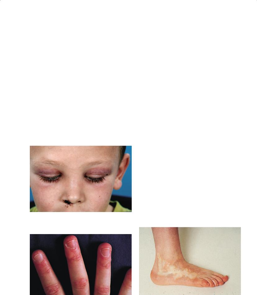
Juvenile dermatomyositis—Children with juvenile DMS may present insidiously and are often labelled as malingerers before the true nature of the diagnosis is appreciated. Typical symptoms include malaise, progressive proximal muscle weakness and muscle pain or discomfort. Arthritis is found in 20% of the patients. Dermatological manifestations include heliotrope rash (purplish discoloration and oedema of the upper eyelids) (Figure 15.1) and malar rash travelling down the naso-labial folds (malar rash in SLE spares the naso-labial folds), Gottron’s papules over the metacarpophalanges, elbows and knees (often mistaken for eczema) and cuticle hyperaemia (due to distended nailfold capillaries) (Figure 15.2). The diagnosis of juvenile DMS is usually made on the basis of typical rash and the finding of symmetrical proximal muscle weakness. Elevated serum muscle enzymes (CPK, aldolase, AST, ALT, LDH) confirm the presence of muscle inflammation. If the child has a typical rash but normal strength and enzyme levels, magnetic resonance imaging (MRI) of a large muscle demonstrates
Figure 15.1 Heliotrope rash in a boy with dermatomyositis
otherwise subtle inflammation on T2 images. In very weak children, respiratory failure and aspiration pneumonia may be life-threatening.
Localized scleroderma syndromes—Most children with scleroderma have a localized disorder characterized by areas of oval (morphea) or linear lesions that traverse over joints, face (coup de sabre deformity) and trunk (Figure 15.3). Some children have frank arthritis and occasionally may develop extensive joint limitations that mimic polyarthritis but produce paucity of inflammatory signs, except morning stiffness.
Systemic vasculitis syndromes such Wegener’s granulomatosis, polyarteritis nodosa or Churg–Strauss syndrome are extremely rare in childhood, and arthritis is not a common feature.
Acute inflammatory conditions
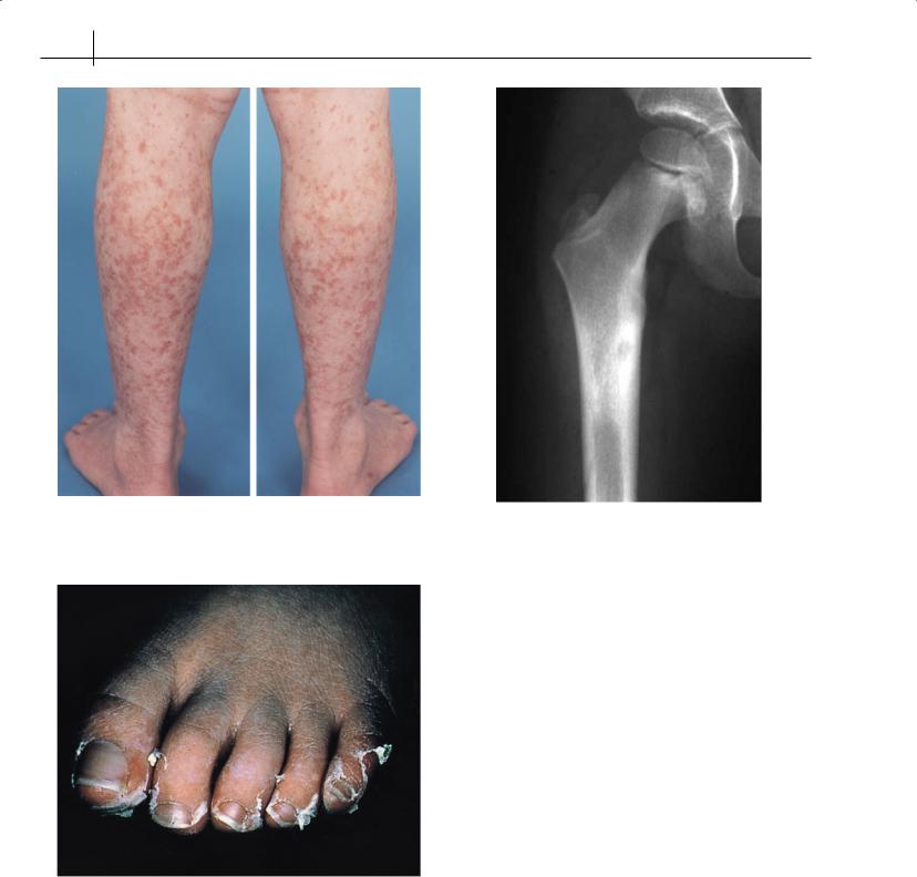
Henoch–Schönlein purpura—HSP is the most common vasculitis of childhood, manifested by purpuric rash over the lower legs and buttocks (Figure 15.4), often associated with cramping abdominal pain, bloody stools, haematuria and occasionally with arthritis of the ankles or knees. Haematuria is virtually always present, and proteinuria may be found, but significant renal disease is extremely rare.
Kawasaki disease—Arthritis is an uncommon feature at presentation in this illness of infants and toddlers; however, atypical cases of Kawasaki disease with fever, rash and elevated inflammatory markers, but without red eyes, raises the suspicion of systemic arthritis (Figure 15.5).
Malignancy
Acute lymphoblastic leukaemia—This may present with bone pain in children (sometimes primarily at night) and even frank arthritis, which can affect one, or sometimes more, joint(s). These children appear toxic and have pain over the affected bone(s). When these children present to the paediatric rheumatologist, their complete blood counts are normal, and a high level of suspicion is required. There is usually anaemia and elevated ESR. Bone-marrow biopsy confirms the presence of immature cells.
Figure 15.2 Gottron’s papules in a boy with juvenile dermatomyositis |
Figure 15.3 Localized scleroderma of the foot of an 11-year-old girl |
90 ABC of Rheumatology
Figure 15.4 Leg purpura of Henoch–Schönlein purpura |
Figure 15.6 Benign tumours, such as osteoid osteoma, may present with |
|
|
|
night pain in the affected limb. These conditions should be considered in |
|
the assessment of a child with “growing pains” |
Figure 15.5 Desquamation of toes in a child with Kawasaki disease
Neuroblastoma—Neuroblastoma is a particularly concerning possibility in younger children who present with fever and joint pain. Early metastases to the bone cause pain that may be difficult for the doctor to localize.
Lymphoma—Lymphoma usually affects older children and may present with musculoskeletal symptoms.
Primary bone malignancies—These are rare and usually visible on plain X-ray radiographs (Figure 15.6).
Idiopathic pain syndromes
The most dramatic musculoskeletal pain in children is often found in the idiopathic pain syndromes. These may be localized (complex regional pain syndromes) or generalized (diffuse pain syndromes, also called fibromyalgia). Exogenous stress (including school pressures, bullying or other forms of abuse, and even parental pressure) is a common accompanying feature, although often unrecognized by the parents. These children and adolescents deserve meticulous physical examination and judicious investigation to rule out an underlying organic pathology. Too much investigation and vacillating doctor-to-patient communication, however, may perpetuate or exacerbate the clinical features of these disorders. Indeed, treatment should start while investigations are ongoing, because disability is common. Occasionally, an idiopathic pain syndrome may complicate a pre-existing condition, such as juvenile idiopathic arthritis. An individualized, intensive, multi-professional, rehabilitation regimen, either in the community or on an inpatient basis, is essential to restore function.
Complex regional pain syndromes—Previously called reflex sympathetic dystrophy, complex regional pain syndromes may begin after trauma (often minor) or without a clear precipitant. They are always associated with immobility, followed by increasing pain,

Juvenile Idiopathic Arthritis |
91 |
|
|
|
|
hypersensitivity, cool skin and complete refusal to use the affected area (Box 15.5). Invariably, patients rate their pain as 9/10, whereas children with JIA typically rate their pain at 2–3/10. There is complete lack of correlation between physical findings and the patient’s level of pain and disability. This condition seems to be increasing in frequency and affecting younger children.
Diffuse musculoskeletal pain syndromes—These poorly defined conditions are characterized by disturbed sleep patterns (initial insomnia, exhausted awakening and napping during the day), tenderness over soft-tissue “trigger” points (with facial grimacing and a sharp intake of breath), and the absence of other findings to suggest organic disease.
Investigations in children with arthritis
JIA is a label for children who fulfil classification criteria made on clinical features alone, as no diagnostic tests exist. Investigations are thus aimed at excluding a wide range of differential diagnoses. However, certain classic patterns emerge as features of history and physical examination combine with typical laboratory and imaging findings, allowing the clinician to arrive at the correct diagnosis.
Haematology
Complete blood count—To look for leucopenia, Coombs’ positive anaemia and thrombocytopenia of SLE and MCTD, as well as the elevated white blood cell (WBC) count and anaemia of chronic inflammation of systemic arthritis. Malignancy may not be ruled out without bone-marrow biopsy.
Erythrocyte sedimentation rate—ESR is elevated in systemic arthritis and SLE but normal in up to half of patients with JIA and in most children with dermatomyositis.
Bone-marrow aspirate—To exclude malignancy, especially before instituting corticosteroid treatment.
Biochemistry
C-reactive protein—C-reactive protein (CRP) is elevated in systemic arthritis but normal in up to half of patients with JIA, as well as patients with SLE, MCTD and DMS.
Urinary catecholamines—To exclude neuroblastoma.
Box 15.5 Features of complex regional pain syndrome
Characteristic
•Severe pain out of proportion to physical findings
•Hyperaesthesia
•Allodynia
•Immobility of affected limb, including adopting a bizarre posture or gait
Occasional
•Limb swelling
•Mottling of skin
•Cool pallor of skin
Liver function tests, creatinine kinase, aldolase, LDH—To rule out dermatomyositis.
Urinalysis—To rule out renal disease of SLE and HSP.
Immunology
Antinuclear antibodies—Positive in low titre in most children with JIA (especially oligoarthritis), and in up to 15% of the general paediatric population. High positive ANAs are seen in virtually all children with SLE and MCTD.
Rheumatoid factor—Negative in 95% of children with JIA but present in 25% of children with SLE.
Immunoglobulins—One in 500 children with JIA has a low level of immunoglobulin A (IgA). IgG is highly elevated in SLE.
Antistreptolysin “O” titre and viral serology—To help with acute rheumatic fever, post-streptococcal arthritis, viral and post-viral conditions.
Borrelia burgdorferi serology—To rule out Lyme disease if there is a history of travel in endemic areas.
Synovial fluid analysis—This is mandatory in suspected septic arthritis, but does not help in other differential diagnoses. Children do not get gout.
Radiology
Plain X-ray radiographs—To rule out fractures, avascular necrosis of bone, bone neoplasia, bone dysplasia and osteomyelitis.
Ultrasonography—To confirm the presence of joint effusion; to look for neuroblastoma.
Technetium-99 bone scan—To highlight bony inflammation secondary to infection, malignancy or benign tumours such as osteoid osteoma.
MRI of joints—To confirm the presence of early synovial inflammation and early erosions; to confirm the presence of myopathy; to rule structural abnormalities.
Classification of JIA
See Box 15.6 for a classification of JIA by the International League of Associations for Rheumatology.
Box 15.6 International League of Associations for
Rheumatology classification of juvenile idiopathic arthritis
•Systemic arthritis
•Oligoarthritis—persistent
•Oligoarthritis—extended
•Polyarthritis—rheumatoid-factor negative
•Polyarthritis—rheumatoid-factor positive
•Psoriatic arthritis
•Enthesitis-related arthritis
•Undifferentiated arthritis