Материал: ABC-of-Rheumatology-ABC-Series-
82 ABC of Rheumatology
Box 14.4 Patterns of psoriatic arthritis
• Asymmetrical oligoarthritis (50%); involvement of one to five joints
• Predominantly distal interphalangeal joint disease (5–10%); distinctive but unusual form of psoriatic arthritis
• Rheumatoid pattern (25%); symmetrical small-joint arthritis particularly affecting metacarpophalangeal, wrist and proximal interphalangeal joints; may be indistinguishable from rheumatoid arthritis
• Arthritis mutilans (1–5%); osteolysis results in destruction of the small joints of the digits with shortening
• Spondyloarthritis (20%); may be isolated sacroiliitis, atypical or typical AS
*
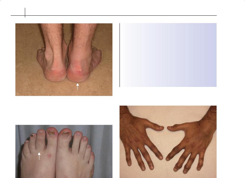
Figure 14.3 Achilles tendon bursitis
Figure 14.4 “Sausage toe” |
Figure 14.5 Distal interphalangeal joint involvement in psoriatic arthritis |
The clinical course and disease severity of AS are highly variable. Inflammatory back pain and stiffness dominate the picture in the early stages, whereas chronic pain and deformity may develop over time. Osteoporosis tends to develop early in the disease, predisposing to spinal fractures later. One-third of AS sufferers give up work before retirement age because of their disease.
Psoriatic arthritis
Psoriatic arthritis is an inflammatory arthritis associated with psoriasis, usually with negative tests for rheumatoid factor. It is not a homogeneous clinical entity. In common with other spondyloarthritides, the key features are seronegative arthritis, enthesitis and, in a minority, sacroiliitis or spondylitis. It is the only SpA in which small joints of the hand are frequently affected. Five patterns of joint involvement are recognized (Box 14.4), although many patients have overlapping patterns of disease.
Psoriasis occurs in 5% of most white populations, and 5–15% of sufferers develop one or another form of associated arthritis. In a small minority of patients arthritis precedes the onset of psoriasis.
Typical psoriatic nail changes such as pitting, onycholysis and hyperkeratosis are seen in over 80% of patients with psoriatic arthritis, although skin lesions may be subtle and should be sought specifically in the scalp and natal cleft. Arthritis is characteristically oligoarticular and asymmetrical and may be associated with dactylitis of fingers or toes, often described as a “sausage digit” (Figure 14.4). Distal interphalangeal joint involvement at the fingers is uncommon but highly characteristic (Figure 14.5). Enthesitis plays a role in dactylitis but may also occur at more typical sites around the patella or around the heel at the Achilles tendon or plantar fascia insertion. Twenty per cent of patients with psoriatic arthritis develop low back pain with sacroiliitis and may develop typical or atypical spondylitis. Conjunctivitis and anterior uveitis may occur but less commonly than in AS.
Spondyloarthritides 83
Reactive arthritis
Reactive arthritis (ReA) is aseptic arthritis that occurs subsequent to an extra-articular infection, typically of the gastrointestinal (GI) or genito-urinary (GU) tract. The key GI pathogens are Salmonella typhimurium, Yersinia enterocolitica, Shigella flexneri and Campylobacter jejuni; the commonest GU pathogen is Chlamydia trachomatis. The true incidence and prevalence of ReA are not well defined. In epidemics involving Salmonella or Yersinia, ReA develops in up to 7% of infected individuals, but in as many as 20% of B27-positive individuals. In such epidemic studies, B27 confers risk not only for the onset of arthritis but also for axial involvement and chronicity. Typically, arthritis begins 1 to 3 weeks after the GI or GU infection.
As with other SpA syndromes, the pattern of joint involvement in ReA is one of asymmetrical oligoarthritis mainly affecting joints of the leg. As in AS, enthesitis may arise as Achilles tendonitis or plantar fasciitis, and dactylitis may occur at one or more toes. Sacroiliitis, with buttock pain, may occur in the acute phase, but radiographic changes are seen largely in the patients with a chronic course.
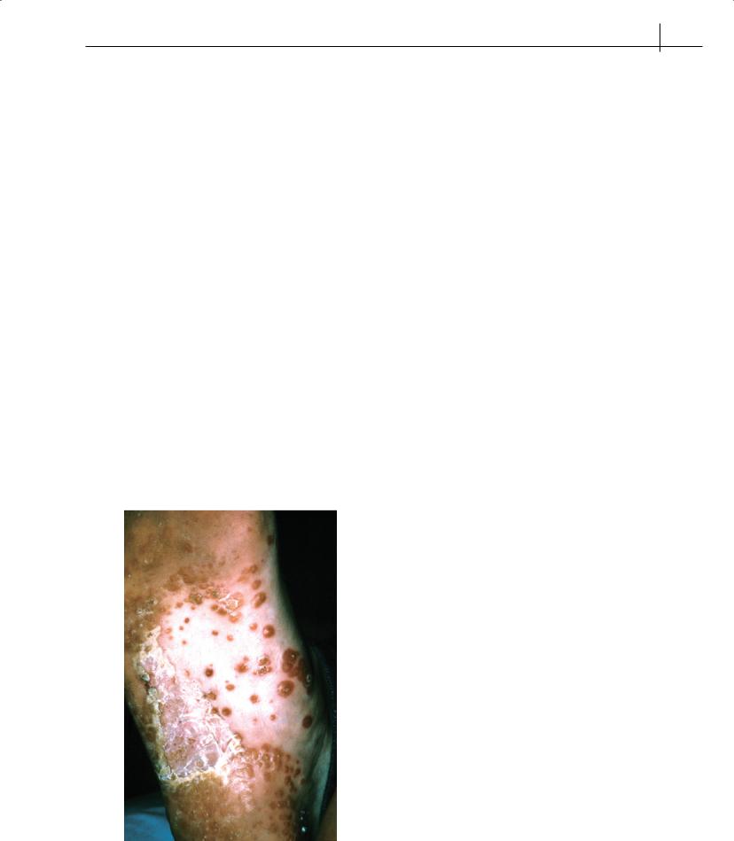
When ReA is accompanied by urethritis, conjunctivitis or mucocutaneous lesions, the term “Reiter’s syndrome” may be applied, but increasingly ReA is used to refer to this symptom complex. Urethritis may be manifest as dysuria or discharge and psoriasiform skin, and mucosal lesions include circinate balanitis and keratodermablennorrhagicum(Figure14.6),apainlesspapulosquamous
Figure 14.6 Keratoderma blennorhagicum
eruption on the palms or soles. Painless lingual or oral ulcers may also be seen. Conjunctivitis is usually bilateral and painful. Acute anterior uveitis is usually unilateral and may not be synchronous with the acute episode but may be clinically indistinguishable from conjunctivitis.
The most important differential diagnosis for ReA is septic arthritis, so appropriate culture of synovial fluid should precede the diagnosis of ReA whenever possible. The course of ReA is variable, and few prognostic markers are available for the clinician to predict the course in any individual case. The majority of patients have an initial episode lasting 2 to 3 months, but synovitis may persist for a year or longer. In patients with chronic disease a significant minority develop some degree of functional disability.
Enteropathic arthritis
Enteropathic arthritis is an inflammatory arthritis associated with inflammatory bowel disease (IBD), particularly ulcerative colitis and Crohn’s disease. Two patterns of peripheral joint involvement are recognized, designated type 1 and type 2. Usually bowel and joint symptoms occur independently, and arthritis may wax and wane over many years. Non-specific arthralgia and myalgia without an inflammatory component, similar to that seen in fibromyalgia, is not uncommon in people with IBD.
Type 1 arthropathy affects approximately 5% of patients with IBD. Typically the peripheral arthritis is oligoarticular and principally affects the knees. It is usually self-limiting without leading to joint deformities. Joint symptoms can occur early in the course of bowel disease and may precede the onset of bowel symptoms. Enthesitis of the Achilles tendon and plantar fascia and dactylitis may also occur.
Type 2 arthropathy affects approximately 3% of patients with IBD. Arthritis is usually polyarticular, principally affecting the metacarpophalangeal joints, although the knees, ankles, elbows, shoulders, wrists, proximal interphalangeal joints and metatarsophalangeal joints may also be affected, sometimes in a migratory fashion.
Sacroiliitis and spondylitis occur in up to 20% of patients with either form of IBD. The course of spinal involvement is completely independent of the course of the IBD and may precede by years the first manifestations of bowel disease.
Undifferentiated spondyloarthritis
The development of inflammatory back pain or peripheral large joint arthritis, often in individuals who are positive for HLA-B27, with or without other features of the seronegative SpA but without fulfilling criteria for any particular subtype, is referred to as undifferentiated SpA. Most patients are young adults, although children may be affected; a proportion of cases will evolve over time to into a classifiable subset, particularly ankylosing spondylitis.
It is not unusual for the first feature of a spondyloarthritis to be an enthesitis, especially at the Achilles tendon or plantar fascia. These lesions may also occur independently of any arthritic conditions, especially in athletes. In spondyloarthritis, Achilles tendonitis typically affects the actual entheseal junction, often with marked

84 ABC of Rheumatology
bone oedema visible on MRI scanning and sometimes with Achilles tendon bursitis; in athletes pain and tendon swelling occur higher up in the tendon close to the muscle belly. Plantar fasciitis is not so easily differentiated, although it often occurs in overweight older adults.
There are no diagnostic criteria for undifferentiated SpA per se; however, the European Spondyloarthropathy Study Group (ESSG) has set out criteria, as in Box 14.1.
Treatment
The goals of treatment are to relieve symptoms, improve function and delay or prevent structural damage. To some extent treatment of spinal inflammation differs from that of peripheral joint synovitis and enthesitis, so treatment must be tailored to the actual problems in the individual patient at the time.
Sacroiliitis and spondylitis
First-line treatment—Regular physiotherapy and encouragement to exercise regularly; use of non-steroidal anti-inflammatory drugs (NSAIDS), such as naproxen or diclofenac, or COX-2 inhibitors such as etoricoxib or celecoxib.
Second-line treatment—Oral and intramuscular corticosteroids may control spinal symptoms, but long-term use should be avoided; local corticosteroid injections into one or both sacroiliac joints under radiographic imaging may be helpful.
Third-line treatment—Anti-tumour necrosis factor agents have been proven significantly to reduce spinal pain and stiffness and to increase spinal movement and patient function; reduction of spinal inflammation has been demonstrated by MRI scanning.
Adjunctive treatment—Bisphosphonates, calcium and vitamin D may improve bone density, although fracture reduction has not been demonstrated in AS; low-dose amitriptyline at night may improve sleep and reduce pain and fatigue.
Oligoarthritis and/or enthesitis
First-line treatment—Analgesics such as paracetamol/acetaminophen or codeine-based drugs may be helpful; intra-articular or intra-lesional corticosteroid injection can be useful for single peripheral joint involvement or enthesitis; injections into weightbearing tendons should be avoided; NSAIDs may provide symptomatic relief (they must be used with caution in patients with IBD,
as they may exacerbate the gut disease); orthotics, including heel pads, and carefully chosen footwear, including suitable trainer/ running shoes, may provide the best symptomatic relief for arthritis or enthesitis affecting the feet; in patients with ReA, genital tract infection should be treated as in uncomplicated infection, with treatment of sexual contacts; antimicrobial treatment of gut infection is not usually indicated, and there is no clear evidence for long-term antimicrobial treatment of established arthritis.
Second-line therapy—Disease-modifying anti-rheumatoid drugs (DMARDs) may be effective for those patients with aggressive, erosive or polyarticular disease, as in the treatment of rheumatoid arthritis; methotrexate may be effective for both skin and joint disease, although the published evidence is scant; in individuals with enteropathic arthritis, sulphasalazine may be effective for both joint and bowel disease; rigorous monitoring of DMARD therapy, according to established practice guidelines, should be undertaken; oral or intramuscular corticosteroid treatment may be effective, especially in those with marked systemic features; in psoriatic arthritis, steroid therapy carries a risk of an exacerbation of skin disease on steroid withdrawal.
Third-line treatment—In those patients who have an inadequate response to conventional DMARDs and in whom the diagnosis is well established, anti-tumour necrosis factor therapy with etanercept, infliximab or adalimumab may be dramatically effective in the control of joint and skin disease; etanercept has not been shown to be effective for IBD.
Further reading
Carlin EM, Keat AC. European guidelines for the management of sexually acquired reactive arthritis. International Journal of STD and AIDS 2001; 12
(Suppl. 3) 94–102.
Hannu H, Inman RD, Granfors K, Leirisalo-Repo M. Reactive arthritis or postinfectious arthritis. Best Practice and Research. Clinical Rheumatology
2006; 20: 419–433.
Holden W, Orchard T, Wordsworth P. Enteropathic arthritis. Rheumatic Diseases Clinics of North America 2003; 29: 513–530.
Rudwaleit M, Metter A, Listing J, Sieper J, Braun J. Inflammatory back pain in ankylosing spondylitis: a reassessment of the clinical history for application as classification and diagnostic criteria. Arthritis and Rheumatism 2006; 54; 569–578.
Zochling J, van der Heijde D, Burgos-Vargas R et al. ASAS/EULAR Recommendations for the management of ankylosing spondylitis. Annals of the Rheumatic Diseases 2006; 65: 442–452.

CHAPTER 15
Juvenile Idiopathic Arthritis
Taunton R Southwood1 and Ilona S Szer2,3
1University of Birmingham, and Birmingham Children’s Hospital NHS Foundation Trust, Birmingham, UK
2UCSD School of Medicine, San Diego, CA, USA
3University of California, San Diego, CA, USA
OVERVIEW
•Juvenile idiopathic arthritis (JIA) is not the same disease as rheumatoid arthritis.
•JIA is an umbrella term that includes at least seven different conditions, each representing a unique form of childhood arthritis.
•JIA is characterized by persistent, non-recurrent, objective joint swelling.
•Joint pain and stiffness are presenting symptoms in most, but not all, children and young people with JIA.
•Early diagnosis is the key to optimal outcome.
•Effective treatment is available and positively affects long-term outcome.
Introduction
The diagnosis of arthritis or other rheumatic conditions in children requires an awareness of the age-dependent manifestations of such conditions, skill in history taking, a meticulous approach to physical examination and judicious use of investigations. Such conditions may present with relatively common, non-specific, “constitutional” paediatric symptoms such as fever, rash, fatigue, weakness, anorexia and pain. Individually, or in combination, these are most likely to be features of common, insignificant, transient illnesses. Rheumatic diseases usually have additional clues, albeit subtle ones, that should alert the clinician to a possible rheumatic diagnosis. In arthritic disorders, joint swelling is the pivotal feature, but others include characteristic “rheumatic patterns” of fever, rash, weakness, diurnal variation or disease progression despite simple measures.
The key signs on physical examination may also be subtle, requiring experience and skill to discern and interpret them. A thorough physical examination (including detailed musculoskeletal assessment) of any child with potential rheumatic symptoms is essential. Joint swelling and pain with movement usually confirm the presence of arthritis (note that isolated joint pain/joint tender-
ABC of Rheumatology, 4th edn. Edited by Ade Adebajo. ©2010 Blackwell Publishing Ltd. 9781405170680.
Box 15.1 Criteria for the diagnosis of juvenile idiopathic arthritis
All three conditions must be met:
•Arthritis persisting for >6 weeks
•Onset before age 16 years
•Exclusion of other conditions associated with or mimicking arthritis
ness without swelling are features of arthralgia). Investigations, especially during the first few weeks of illness, are aimed at ruling out the long list of conditions that comprise the differential diagnosis of childhood arthritis.
Juvenile idiopathic arthritis (JIA) is the most likely diagnosis in children with persistent symptoms and clinical features of arthritis for at least 6 weeks (Box 15.1), as most other illnesses either resolve or are treated and referred to other specialists during this time period. For the experienced paediatric rheumatologist, the child with arthritis or other chronic rheumatic illness presents with an almost instantly recognizable pattern of symptoms. The lack of timely detection of childhood inflammatory disease and delay in treatment may adversely affect the course and outcome of JIA. There are also dangers inherent in trying to make the diagnosis of JIA too precipitously, without ruling out other rare but important disorders, such as septic arthritis or malignancy. Diagnostic imaging and laboratory investigation must always be carefully considered,butthereisnopathognomonictestforJIA.Disproportionate over-investigation may increase child and family anxiety without adding value to the diagnostic process. The overzealous investigator may even exacerbate the severity of some conditions, such as chronic idiopathic pain syndromes.
This chapter aims to give an overview of JIA, including clinical features, differential diagnosis, investigations, natural history and principles of treatment. No substitute exists, however, for actual clinical experience, and the reader is strongly recommended to practise the skills of paediatric musculoskeletal examination at every appropriate opportunity. An appreciation of the range of normality in children and young people is an absolute prerequisite to the detection of abnormality.
85

86 ABC of Rheumatology
Clinical features of JIA
JIA is one of the most common physically disabling conditions of childhood, with a prevalence of approximately one in a thousand children under the age of 16 years (surveys suggest that this may be higher, even as many as 4 in 1000 children; 10,000–40,000 affected children in the UK and up to 300,000 in the USA). The incidence of JIA is 1 in 10,000. In typical general practice, however, JIA is rare; one new case may be seen every 20 years. It is difficult to maintain a high index of suspicion for JIA in the face of this degree of rarity.
Most affected children are in their preschool or early school years, and often have difficulty describing their symptoms. Parents may notice joint swelling if one or more large peripheral joints are involved, such as the knee (the most common joint affected), ankle or wrist. It is rarer for children to present with isolated small joint (finger or toe) arthritis or axial joint involvement (such as the shoulder, hip, spine or temporomandibular joints), and parents are also less likely to notice swelling in these joints. Diurnal variation of symptoms, such as early morning joint stiffness or exacerbation after prolonged rest (joint “gelling”) are characteristic. Stiffness improves with movement and may be helped by a warm bath or shower. Duration of morning stiffness may provide an index of improvement with treatment. Joint dysfunction may be manifest by limping, difficulty with writing or inability to carry out other activities of daily living (Table 15.1).
There is accumulating evidence that the natural history of JIA may not be as benign as first thought; between a third and a half of patients have persistent joint inflammation into their adult years. More aggressive treatment is being used in an attempt to
Table 15.1 Typical symptoms of juvenile idiopathic arthritis
Present in all forms of JIA
Joint symptoms (pain, dysfunction, stiffness), particularly after sleep or prolonged sitting
Persistent joint swelling, particularly of the knee, ankle, wrist and small joints of the hand
Difficulty chewing, asymmetric mouth opening and micrognathia
Muscle atrophy
Flexion contracture deformity
Synovial hypertrophy
Constitutional symptoms (dramatic in children with systemic arthritis but mild in polyarthritis and ERA)
Fever (high and quotidian in systemic arthritis, low grade in polyarthritis, no pattern in ERA)
Rash (maculopapular, recurrent in systemic arthritis, nodules in polyarthritis, rheumatoid factor+, psoriasis and nail pitting in psoriatic arthritis)
Growth failure (almost always severe in systemic arthritis, mild in polyarthritis and localized to specific bones in oligoarthritis)
ERA = enthesitis-related arthritis
induce early disease remission, an approach that has been complemented recently by a wider therapeutic armamentarium. Several targeted biologic drugs have been shown to be beneficial in JIA.
Physical findings in JIA
Peripheral joint arthritis in children is usually accompanied by joint swelling. In the knee, this may be demonstrated by balloting synovial fluid, palpating a fluid thrill on joint movement, eliciting a positive patella tap, or occasionally finding a Baker’s cyst in the popliteal fossa. Swelling of the ankle may distort the contours of medial or lateral malleoli. When the ankle is dorsiflexed, the usually prominent anterior tendon surface markings may be obscured by arthritis, although this may be difficult to see in infants and overweight children. Other relevant observations include muscle wasting, particularly of the vastus medialis and gastrocnemius, and leg length discrepancy, which often indicates accelerated growth around affected joints.
Wrist arthritis may be best appreciated by asking the child to press the palms of their hands together in the “prayer” position; a dorsal bulge and reduced range of movement, especially if it is asymmetrical, are consistent features of synovitis. Swelling of the elbow can be palpated on either side of the olecranon and usually results in a flexion deformity of the elbow. Elbow swelling obscures the posterior dimple created when the elbow is fully extended.
The small joints of the hands and feet should be inspected and palpated individually; reliable signs of synovitis are the presence of joint margin tenderness, restricted movement, swelling and purplish discoloration, incomplete fist closure and diminished grip strength. Cervical spine involvement may be detected by inability to rotate the head laterally to place the chin on each shoulder and by reduced cervical extension. Temporomandibular synovitis is often missed; it may prevent full and symmetrical opening of the mouth. Involvement of sacroiliac joints and low back in patients with enthesitis-related arthritis (ERA) can be documented by a limited modified Schober test (less than 6 cm expansion of lumbar spine with forward bending) and documentation of tenderness of sacroiliac joints to direct palpation. Enthesopathy, a hallmark of ERA, is found at tendon insertions into bones, most frequently where the Achilles tendon inserts into the calcaneus. Careful observation of gait allows the examiner to evaluate the function of lower limb joints.
Children with JIA have a variety of extra-articular physical findings that help establish the diagnosis. For example, children with systemic arthritis may have little in the way of articular signs initially, but other characteristic features may be prominent, including a pink, macular, truncal rash (which may be pruritic and exhibit Koebner’s phenomenon), lymphadenopathy, hepatosplenomegaly and myalgia. Children with oligoarthritis tend to appear very healthy and have few findings aside from arthritis (most frequently the knee). If asymptomatic chronic anterior uveitis has preceded the onset of arthritis, posterior synechiae and/ or band keratopathy may be visible with a hand-held ophthalmoscope focused on the lens.
Once the presence of arthritis is confirmed objectively, it is vital to exclude conditions that may mimic JIA. Most serious among