Материал: ABC-of-Rheumatology-ABC-Series-
72 ABC of Rheumatology
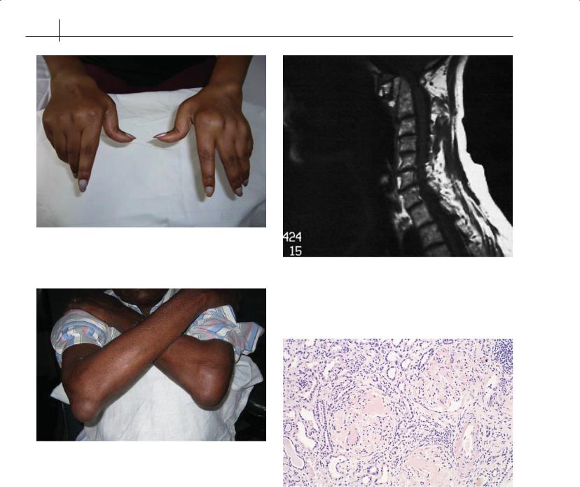
Figure 12.1 Typical changes in the hands in rheumatoid arthritis
Figure 12.2 Large rheumatoid nodules over the elbows
“Red flags”
A variety of complications of RA or its treatment can occur and require vigilance on the part of clinicians to pick them up early and intervene to prevent severe morbidity, and even mortality in certain cases; some of these are detailed below.
Atlanto-axial subluxation—This results from involvement of the atlanto-axial joint, which may be clinically asymptomatic until the subluxation develops. Development of pain around the occiput, radiating arm pain, numbness or weakness of the limbs and vertigo on neck movement are warning signs; if not picked up this may lead to sudden death, especially if patients undergo neck manipulation for endotracheal entubation during surgical procedures. It is advisable to actively look for it as part of pre-surgical evaluation. It can be picked up easily by doing lateral views of the cervical spine
Figure 12.3 Magnetic resonance image of the cervical spine showing atlanto-axial involvement in rheumatoid arthritis
Figure 12.4 Renal biopsy showing amyloid deposit (Congo Red stain)
in flexion (Figure 12.3) and extension and measuring the distance between the posterior margin of the atlas ring and the anterior surface of the odontoid process.
Amyloidosis—Renal deposition of amyloid (Figure 12.4) is a recognized feature of longstanding RA and should be suspected if the patient develops increasing leg oedema, proteinuria and worsening renal functions. Drug-related causes such as goldor penicilla- mine-induced proteinuria need to be ruled out. A renal biopsy will conclude the diagnosis.
RA: Clinical Features and Diagnosis |
73 |
|
|
|
|
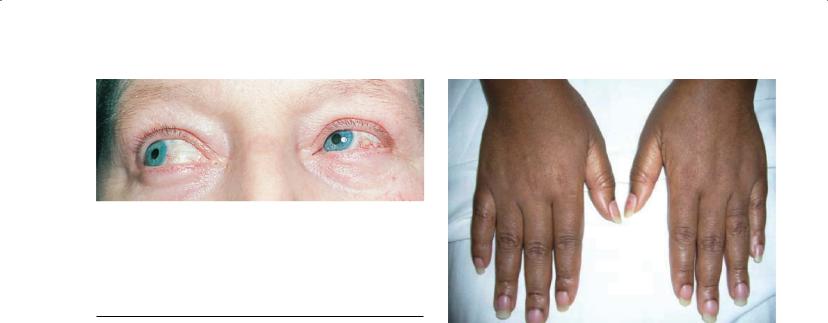
Figure 12.5 Epicleritis in rheumatoid arthritis
Table 12.1 Other manifestations of rheumatoid arthritis
Haematological |
Cutaneous |
Anaemia |
Rheumatoid nodules |
Neutrophillia |
Peripheral vasculitis |
Thrombocytosis |
Leg ulcers |
Felty’s syndrome |
Alopecia |
Neurological |
Ocular |
Entrapment neuropathies such |
Xeropthalmia |
as carpal tunnel syndrome |
|
Mononeurits multiplex |
Scleritis |
Peripheral neuropathies |
Episcleritis |
Pulmonary |
Others |
Pleural effusions |
Dry mouth |
Interstitial lung disease |
Osteoporosis |
Bronchiolitis oblitrans |
Muscle wasting |
Cardiac |
|
Pericarditis |
|
Coronary vasculitis (rare) |
|
|
|
Pericarditis—Onset of central chest pain worsened by lying flat, accompanied by a pericardial rub, merits urgent echocardiogram to confirm and urgent initiation of steroid therapy. Infective causes such as tuberculosis need to be ruled out by aspiration and analysis when suspected.
Monoarticular flare—A single joint worsening should always be viewed with suspicion in RA, and septic arthritis needs to be looked for. It is prudent to initiate treatment for possible septic arthritis until the results of the joint aspirate rule it out.
Eye involvement—Sudden onset of eye pain and increased lacrimation should alert the clinician to the possibility of scleritis (Figure 12.5); if left untreated this may lead to full-thickness involvement of the sclera, with thinning and risk of perforation. Called scleromalacia perforans, this sinister condition is thankfully rare but needs to be looked out for.
Other manifestations of RA are given in Table 12.1.
Figure 12.6 Subtle features of synovitis in early rheumatoid arthritis
Diagnosis
The diagnosis of RA is predominantly a clinical one; no diagnostic test has been shown to be foolproof, and both false-positive and false-negative results are seen with varying frequency.
History
A detailed history of the problem, its onset and progression with time, relieving and aggravating factors and the distribution of the symptoms are all important elements in the history. A progressive pattern of joint involvement, stiffness and increased pain after a period of inactivity and a history of joint swellings are indicative of inflammatory joint disorders. A family history of rheumatological disease can raise the suspicion further. The distribution of joint involvement helps in distinguishing other forms of arthritides such as spondyloarthritis and psoriatic arthritis.
Clinical examination
The objective of the clinical assessment is to identify signs of inflammatory arthritis, such as swelling, tenderness and restriction of movement of the joints. A symmetrical involvement of the hands, especially the metacarpophalangeal and proximal interphalangeal joints, with relative sparing of the axial skeleton, are some key elements that support the diagnosis of RA. Clinical evaluation may also pick up extra-articular findings that can support the diagnosis or refute it—for example, the presence of rheumatoid nodules and psoriatic skin patches, respectively. In early disease the classical signs of structural changes may be missing and subtle synovitis (Figure 12.6) may escape notice; however, tenderness and restriction without history of trauma should arouse suspicion.
Laboratory evidence
Active RA is associated with a variety of haematological responses. Acute-phase responses such as a high erythrocyte sedimentation rate or C-reactive protein, a high platelet count and high serum
74 ABC of Rheumatology
Box 12.1 Other causes of positive rheumatoid factor
•Other connective tissue diseases
•Viral infections
•Leprosy
•Tuberculosis
•Subacute bacterial endocarditis
•Sarcoidosis
•Liver diseases
•Leishmaniasis
ferritin can be seen in most patients. Anaemia of chronic disease may be present in many patients with chronic conditions. A very high leucocyte response is uncommon and usually indicative of an infection, which should be looked for in such situations.
The traditional test of rheumatoid factor that detects immunoglobulin M (IgM) antibodies directed against IgG can be used as supporting evidence in establishing the diagnosis, but is neither conclusive nor universal in patients with RA. A number of conditions are associated with the presence of rheumatoid factor in serum (Box 12.1).
A new test that seems more promising, called anti-cyclic citrullinated peptide (anti-CCP), is now commercially available. This test seems to be more specific (95–98%) for the diagnosis of RA and it is sensitive (50–60%) in early rheumatoid disease. It is also a marker of erosive disease and can predict eventual development of RA in undifferentiated arthritis.
Radiology
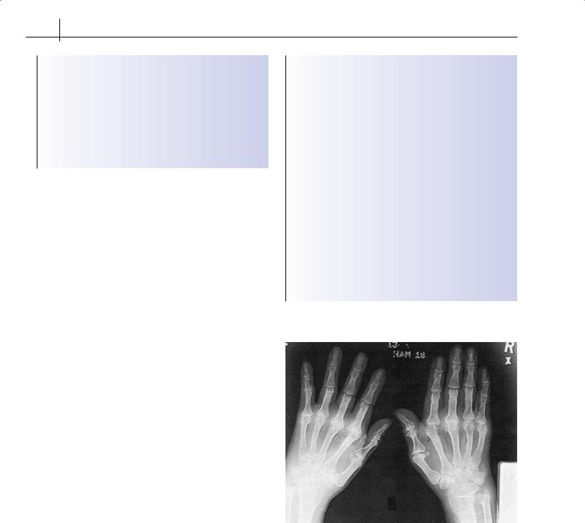
Radiological features of classical periarticular erosions (Figure 12.7) are characteristic and may appear within the first 3 years of disease in the majority of the patients; more subtle changes, such as juxta-articular osteopenia and early joint-space narrowing, are less specific and can be misleading. Conventional radiology can still be useful in monitoring progression of the disease in established diagnosis and to plan corrective surgeries when there is significant disability. Newer modalities such as magnetic resonance imaging are now increasingly employed to detect early synovitis and bone oedema and can be utilized effectively in picking up early disease. Ultrasonography may be useful in picking up joint effusions, a Baker’s cyst and pleural disease. High-resolution computed tomography is the modality of choice in picking up interstitial lung disease and pulmonary fibrosis. It should be carried out in patients with progressive loss of lung function.
Synovial fluid analysis
This test is rarely required to establish diagnosis in a typical presentation; however, in atypical presentations with large joint involvement, especially monoarticular, it is vital to rule out infective aetiology and crystal arthropathy. The fluid would typically show a high protein and leucocyte count and the absence of crystals and organisms on Gram stain.
Box 12.2 American College of Rheumatology revised criteria for the classification of acute rheumatoid arthritis
(1987)
Diagnosis of rheumatoid arthritis needs four of seven of the following criteria. In criteria 1–4, the joint signs or symptoms must be continuous for at least 6 weeks.
1. Morning stiffness of duration longer than 1 hour
2. Arthritis of three or more joint areas simultaneously*
3. Arthritis of hand joints (wrist, metacarpophalangeal joints or proximal interphalangeal joints)
4. Symmetric arthritis* (bilateral involvement of metacarpophalangeal, proximal interphalangeal or metatarsophalangeal joints is acceptable without absolute symmetry)
5. Rheumatoid nodules as observed by the doctor
6. Serum rheumatoid factor, as assessed by a method positive in less than 5% of control subjects
7. Radiographic changes, as seen on anteroposterior radiographs of wrists and hands
*Possible areas: proximal interphalangeal joint, metacarpophalangeal joint, wrist, elbow, knee, ankle and metatarsophalangeal joint (observed by a physician).
Figure 12.7 Radiograph of the hands, showing erosions at the metacarpophalangeal and proximal interphalangeal joints
The American College of Rheumatology has formulated and modified classification criteria to aid in diagnosis of RA (Box 12.2); however, these criteria have poor sensitivity in picking up early rheumatoid disease.
Differential diagnosis
Other arthritides can be distinguished on the basis of jointinvolvement pattern; however, atypical presentations may prove

RA: Clinical Features and Diagnosis |
75 |
|
|
|
|
Figure 12.8 Psoriatic rash at the natal cleft
challenging to rule out. A careful search for evidence of nail pitting or skin lesions may clinch the diagnosis in psoriatic disease (Figure 12.8), but joint aspiration for crystals may be needed to exclude
polyarticular gout. Malignant conditions such as leukaemias and lymphomas should be sought, especially in acute presentations in younger patients. In areas of high incidence, conditions such as hepatitis B and C and HIV need to be borne in mind.
As RA is a chronic disease that leads to significant morbidity and disability, the clinician has the vital responsibility of making an early diagnosis and commencing treatment early to prevent these problems from occurring. No laboratory tests are diagnostic, and ultimately the diagnosis relies on a clinical evaluation by the practitioner.
Further reading
Firestein GS, Panayi G & Wollheim F, eds. Rheumatoid Arthritis, 2nd edn. Oxford University Press, Oxford, 2006.
Isaacs J, Moreland LW. Fast Facts: rheumatoid arthritis. Health Press, Oxford, 2002.
Isenberg D, Maddison P, Woo P, Glass D, Breedveld F, eds. Oxford Textbook of Rheumatology. Oxford University Press, Oxford, 2004.
Taylor P. Rheumatoid Arthritis in Practice. Royal Society of Medicine Press, London, 2006.

CHAPTER 13
Treatment of Rheumatoid Arthritis
Edwin S L Chan1, Anthony G Wilson2 and Bruce N Cronstein1
1New York University School of Medicine, New York, USA
2University of Sheffield, Sheffield, UK
OVERVIEW
•Rheumatoid arthritis (RA) is a disease requiring life-long treatment.
•Treatment should begin early because radiological damage can occur much earlier than previously thought.
•Disease-modifying antirheumatic drugs (DMARDs) and biological-response modifiers have been proven to retard disease progression.
•Combination therapy is often more efficacious than monotherapy.
•Inadequately treated rheumatoid arthritis is associated with increased mortality.
Remarkable strides have been made in controlling clinical and radiological progression of rheumatoid arthritis (RA) in recent years. However, the usefulness of small molecules such as methotrexate has not been overshadowed by the current interest in biological-response modification. Our understanding of the molecular mechanisms responsible for the pathogenesis of RA has heralded a shift from empiricism to selective molecular targeting in immunomodulatory pharmacotherapeutics. While symptomatic control and reduction of the clinical signs of synovitis have been the foremost considerations in the past, modern pharmacotherapy has emphasized the need to slow down, if not halt, disease progression as well as to prevent the development of potential complications. It is now recognized that significant documented radiological damage can occur in this disease much earlier than previously thought, certainly within the first 2 years of disease onset. Disease-modifying therapy is therefore introduced early following confirmation of diagnosis, particularly in those with poor prognostic indicators, such as severe disease activity, radiological damage or anti-cyclic citrullinated peptide positivity. Some favour a more aggressive combination of drugs in early disease, with a
ABC of Rheumatology, 4th edn. Edited by Ade Adebajo. ©2010 Blackwell Publishing Ltd. 9781405170680.
possible “step-down” approach once the disease comes under control. The old “pyramidal” treatment approach has therefore been called into question, and new advances have dramatically improved the disease outlook for the RA patient.
Non-steroidal anti-inflammatory drugs
Non-steroidal anti-inflammatory drugs (NSAIDs) are inhibitors of cyclooxygenase, an enzyme that catalyses the conversion of arachidonic acid to prostanoids. The enzyme exists in two isoforms. Cyclooxygenase-1 is constitutively expressed in many tissues including platelets, blood vessels and the upper gastrointestinal (GI) mucosa, where production of prostaglandin E2 mediates a protective mucosal effect that includes mucus secretion and diminution of acid production. Expression of cyclooxygenase-2 is induced at sites of inflammation, particularly on polymorphonuclear cells and macrophages. Thus, non-selective inhibition of both isoforms by traditional NSAIDs may ameliorate desirable gastroprotective effects mediated by cyclooxygenase-1, and reported hospitalization of RA patients as a result of upper GI complications may exceed 1% of patients treated per year. Selective inhibition of cyclooxygenase-2, however, has met with concerns over potential cardiovascular risks, although recent evidence suggests that this problem is also associated with several traditional NSAIDs. Despite these concerns, NSAIDs continue to be used for symptomatic control in RA, but it must be emphasized that they have little effect in limiting joint damage or radiological progression.
Corticosteroids
The demonstration of the anti-inflammatory efficacy of corticosteroids in RA resulted in the first Nobel Prize awarded for a clinical observation, and 70 years hence, these potent anti-inflammatory agents continue to have an important place in the management of RA. Furthermore, multiple routes of administration, including depot injections (methylprednisolone and triamcinolone acetonide) and local intra-articular injections offer a variety of therapeutic options. Given orally, the onset of action is quick and is therefore useful in relieving symptoms while awaiting the onset of DMARD activity. Lower oral doses have been favoured (prednisolone up to 10 mg/day) owing to fear of suppression of the hypothalamus– pituitary–adrenal axis, and prevention of corticosteroid-induced osteoporosis must be considered in patients receiving these medications long term.
76