Материал: ABC-of-Rheumatology-ABC-Series-
Osteoporosis 67
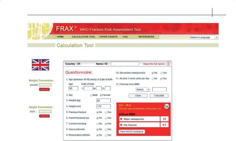
Figure 11.3 The FRAX tool for the assessment of an individual’s 10-year probability of fracture (http://www.shef.ac.uk/FRAX). Once the calculation is completed, clicking on the “View NOGG guidance” button will automatically display the individual’s probability within the suggested care pathways published by the National Osteoporosis Guideline Group (http://www.shef.ac.uk/NOGG)
Assessment of osteoporosis
This largely comprises the assessment of future fracture risk (determines the need for intervention) and the diagnosis or exclusion of underlying causes of osteoporosis.
Assessment of future fracture risk
Clinical risk factors—Several risk factors for fracture have been well established. Many of these risk factors impact on BMD but also contribute independently to future fracture risk. Clinicians frequently take account of these other risk factors in deciding whether treatment is required, and a number of algorithms have been developed to improve the prediction of fracture risk. Recently, the World Health Organization produced an algorithm (FRAX→) that estimates the probability of a major osteoporotic fracture (clinical vertebral, hip, wrist or proximal humerus) or hip fracture alone in the next 10 years (see http://www.shef.ac.uk/FRAX). The algorithm
incorporates BMD as an additional, measurable risk factor to information gleaned from clinical risk factors and adds value to the prediction of risk. BMD is probably of most value in those deemed to be at intermediate or high risk. A history of prior low trauma fracture in adult life is a very important risk factor to identify and can usually be obtained by a good clinical history. In contrast, the suggestion of prior vertebral fracture requires spinal imaging for confirmation and appropriate management. The National Osteoporosis Guideline Group (NOGG) has recently published a new management guideline (Figure 11.3) that integrates FRAX with clinical management algorithms.
Spinal radiographs—Up to half of vertebral fractures are asymptomatic and may be suspected from height loss and the development of kyphosis. The latter features may also result from degenerative spinal disease, however, and radiographs of the thoracic and lumbar spine are important to differentiate fractures from degen-

68 ABC of Rheumatology
erative changes. The importance of vertebral fractures for future fracture risk cannot be overstated, and strategies in the near future will involve the assessment of patients by low-radiation-imaging DXA scans to identify prevalent fractures. In the absence of fractures, the assessment of bone mass on plain radiographs is unreliable, so radiological reports of osteopaenia require confirmation by bone densitometry prior to any therapeutic decisions.
Bone densitometry—After age and prior fragility fracture, BMD is the next major determinant of a person’s risk of fracture. The predictive ability of bone density is comparable with that of blood pressure for determining the risk of cerebrovascular accident and of serum cholesterol for determining the risk of coronary thrombosis. The relative risk of fracture increases approximately 2-fold for each standard deviation decrease in bone density.
BMD is usually measured by DXA—a technique that uses extremely low doses of ionizing radiation to quantify BMD accurately and precisely. DXA of the spine and hip are the optimal clinical measurements for diagnosis. Measurement of bone density in peripheral skeletal sites with techniques such as quantitative ultrasound has useful predictive value for osteoporotic fractures, but appropriate intervention thresholds for these measurements remain uncertain, and they are probably not useful for monitoring responses to treatment.
Currently, no rationale exists for population screening of BMD. If access to bone densitometry is limited, it may be appropriate to treat individuals who have had previous low-trauma fractures or who have other strong risk factors for fracture, such as elderly people who need high-dose corticosteroid therapy. Otherwise, measurements should be targeted to individuals likely to be at increased risk of osteoporosis, where knowledge of BMD will influence management. Traditionally, this has meant the measurement of BMD in all patients with recognized risk factors, an approach encapsulated in the Royal College of Physician Guidelines published in 1999. This guidance has now been updated to incorporate the availability of the FRAX tool for assessing fracture risk (http:// www.shef.ac.uk/NOGG). The National Institute for Health and
Table 11.3 Clinical risk factors used for the assessment of fracture probability
Age |
Secondary causes of |
|
Sex |
osteoporosis including: |
|
Low body mass index (≤19 kg/m2) |
• Rheumatoid arthritis |
|
Previous fragility fracture, particularly of the |
• |
Untreated hypogonadism |
hip, wrist and spine, including morpho- |
|
in men and women |
metric vertebral fracture |
• |
Prolonged immobility |
Parental history of hip fracture |
• |
Organ transplantation |
Current glucocorticoid treatment (any dose, |
• |
Type I diabetes |
by mouth for 3 months or more) |
• Hyperthyroidism |
|
Current smoking |
• |
Gastrointestinal disease |
Alcohol intake of 3 or more units daily |
• |
Chronic liver disease |
|
• |
Chronic obstructive |
|
|
pulmonary disease |
|
Falls* |
|
|
|
|
*Not presently accommodated in the FRAX algorithm, but an important risk factor to be taken into account in patient management
Clinical Excellence (NICE) has also recently published guidance for the primary and secondary prevention of fracture in postmenopausal women (Table 11.3) (http://www.nice.org.uk).
Identifying or excluding underlying causes of osteoporosis
Individuals with a low-trauma vertebral fracture or low BMD for age should be investigated for underlying causes of osteoporosis. In addition to a good clinical history, a small number of investigations can exclude the most common secondary causes of osteoporosis (Box 11.1). Treating the underlying cause often leads to at least partial recovery of bone mass.
Reducing fracture risk
The ultimate goal of osteoporosis management is to reduce the future risk of fracture. This involves educating the patient about the nature of the disease, their fracture risk, lifestyle modification (Box 11.2) and, if necessary, the different types of therapy available.
Box 11.1 Investigations to exclude underlying causes of osteoporosis
Routine
•History and physical examination
•Blood cell count, sedimentation rate or C-reactive protein, serum calcium, albumin, creatinine, phosphate, alkaline phosphatase and liver transaminases
•Thyroid function tests
•Bone densitometry (DXA)
Other procedures, if indicated
•Lateral radiographs of lumbar and thoracic spine/DXA-based vertebral imaging
•Protein immunoelectrophoresis and urinary Bence Jones proteins
•Serum testosterone, SHBG, FSH, LH (in men)
•Serum prolactin
•24-hour urinary cortisol/dexamethasone suppression test
•Endomysial and/or tissue transglutaminase antibodies (coeliac disease)
•Isotope bone scan
•Markers of bone turnover, when available
•Urinary calcium excretion
SHBG = sex-hormone binding globulin; FSH = follicle-stimulating hormone, LH = luteinizing hormone
Box 11.2 Lifestyle modification
Optimizing peak bone mass and reducing bone loss
•Exercise needs to be regular and weight-bearing (such as walking or aerobics); excessive exercise may lead to bone loss
•Dietary calcium may be important, especially during growth
•Avoidance of smoking and excessive alcohol consumption

Osteoporosis 69
In some patients, particularly those with recent vertebral fractures, additional approaches aim to reduce pain and improve mobility.
Guidance
In October 2008, NICE published two Technology Appraisal Guidance documents to address the primary (http://www.nice.org. uk/Guidance/TA160) and secondary (http://www.nice.org.uk/ Guidance/TA161) prevention of osteoporotic fractures with alendronate, etidronate, risedronate, raloxifene, strontium ranelate and teriparatide (secondary prevention only). While welcome, there a number of challenges to the implementation of these guidelines, particularly with regard to prescribing alternative treatments when generic alendronate is contraindicated or not tolerated. NICE plan to have clinical guidelines that will address some of the limitations with these approaches (such as not giving guidance for glucocorti- coid-induced osteoporosis or men). In the meantime, a more pragmatic approach to treatment has been proposed by NOGG with the support of many professional and patient societies. This approach suggests that treatment should be considered when an individual’s probability of fracture is comparable to or exceeds that of a woman of the same age who has already sustained a lowtrauma fracture.
Antiresorptive agents
Bisphosphonates—Alendronate and risedronate are available as once-weekly preparations with evidence for significant reductions in vertebral and non-vertebral fractures. These drugs have largely replaced the use of cyclical etidronate. Ibandronate, available as a once-monthly tablet or a three-monthly intravenous slow injection reduces vertebral fractures with indirect evidence for a reduction in non-vertebral fractures. More recently, zoledronate has become available as a once-yearly short infusion with good evidence of anti-fracture efficacy at all sites. Both NICE and NOGG recommend the use of generic alendronate as first-line therapy, although this may not be suitable for or tolerated by all. Poor absorption of these agents means they must be taken on an empty stomach before breakfast (30 minutes before for alendronate and risedronate; 60 minutes before for ibandronate) or on an empty stomach in the middle of a 4-hour fast (cyclical etidronate). The move away from oral daily dosing regimens to more convenient, less frequent dosing has improved adherence to the bisphosphonates.
Hormone or oestrogen replacement therapy—The use of hormone replacement therapy is no longer thought to be appropriate in the management of osteoporosis unless it is needed to control climacteric symptoms, or in women under 50 who have undergone an early menopause. The safety profile of oestrogen-only therapy appears somewhat better than combined oestrogen–progestogen therapy.
Selective oestrogen receptor modulators—These synthetic agents act as oestrogen agonists on bone and lipids, but without oestrogenlike stimulation of breast and endometrial tissues. Raloxifene reduces the risk of vertebral fracture but has not been shown to decrease the risk of non-vertebral fractures. Like hormone replacement therapy, raloxifene is associated with small increases in the
number of thromboembolic events but conversely is associated with a significant reduction in the number of new cases of breast cancer. Other agents in this class will be available in the near future.
Calcium (1000–1200 mg daily)—This has a less marked effect on fracture reduction than the other antiresorptive agents. Adherence can be problematic, and several preparations are now available to aid patient choice and compliance.
Vitamin D (800 units daily) and calcium (1000–1200 mg daily)—
This has been shown to reduce hip fracture risk in the frail elderly and should be considered in all elderly patients who are housebound or in residential care. In patients at higher risk of fracture, it should be used as adjunctive therapy in combination with another antiresorptive agent.
Calcitonin—Calcitonin may be administered as subcutaneous injections or as a nasal preparation, which is associated with fewer side effects. Calcitonin has been shown to reduce the risk of vertebral fracture. This agent has analgesic properties that may be useful in the acute management of vertebral fracture.
Formation-stimulating agents
Teriparatide and parathyroid hormone—These agents have good evidence for their abilities to increase bone formation (and later bone resorption) with an improvement in bone mass and structure, particularly in trabecular bone such as the vertebrae, with reductions in spine fracture risk. A reduction in non-vertebral fractures has also been shown by recombinant teriparatide (PTH 1-34), possibly mediated by improvements in cortical bone width and/or thickness. They are expensive agents and their use is limited to patients with severe, progressive osteoporosis despite exposure to antiresorptive therapy. Teriparatide is licensed for use in men and women, whereas recombinant parathyroid hormone 1-84 is only licensed for postmenopausal women. Treatment is currently limited to 18–24 month durations and most patients will require treatment with antiresorptive agents after discontinuation to maintain the improvements in bone mass. Very recently, teriparatide has been shown to induce greater increases in spine and hip BMD than alendronate in patients with glucocorticoid-induced osteoporosis and is now licensed for use in this setting.
Alternative agents
Strontium ranelate—Strontium ranelate been shown to significantly reduce vertebral and non-vertebral fracture risk in postmenopausal women. The precise mechanism(s) of action remains unclear, but treatment is associated with significant increases in BMD, partly mediated by the presence of strontium in bone, which impacts on the interpretation of changes in BMD. The change in BMD is therefore a potential marker of adherence to therapy, though it may also complicate future estimates of fracture risk.
Pain relief
Pain relief is frequently adequately achieved with analgesics, but physical measures—such as hydrotherapy or transcutaneous nerve stimulators—may be useful adjuncts to treatment. The pain-

70 ABC of Rheumatology
modulating effects of low-dose antidepressants can be helpful, and many patients benefit from assessment at specialist pain clinics. The pain associated with fractures usually resolves within 6 months, but patients with vertebral fractures may need to be given longterm analgesia because of secondary degenerative disease. NICE has also approved techniques such as vertebroplasty (http://www. nice.org.uk/Guidance/IPG12) and kyphoplasty (http://www.nice. org.uk/Guidance/IPG166) for use in selected patients with recent vertebral fractures and persistent or severe pain. Both techniques give good pain relief. Kyphoplasty may also result in some restoration of vertebral height.
Falls prevention
Predisposing factors, such as postural hypotension or drowsiness due to drugs, should be eliminated where possible. Patients may benefit from physiotherapy to improve their balance and saving reflexes. Patients should be provided with appropriate walking aids, and an environmental assessment should be made of their accommodation to eliminate hazards such as loose rugs and cables. Hip protectors have a limited role to play. Visual assessment and treatment is also important. Assessment via specialized falls clinics may be appropriate, particularly in those individuals with features suggesting a medical cause for falls, such as palpitations or blackouts.
Education
An important part of the management of osteoporosis is education and support of the patient, their carers and their family. Groups such as the National Osteoporosis Society (Box 11.3) have a vital role in this area.
Box 11.3 National Osteoporosis Society (NOS)
Camerton Bath BA2 0PJ
Tel.: 01761 471771 (for general enquiries); 0845 4500234 (for medical enquiries)
Monitoring of treatment
The rationale for monitoring treatment response is that a proportion of patients fail to respond to treatment, commonly due to non-persistence with therapy, poor dosing compliance or, less commonly, due to underlying disease. The current standard measure used to monitor treatment response is spine DXA at 18–24 months after treatment initiation. Biochemical markers of bone turnover may offer a more rapid assessment of treatment response—within 3–6 months. The decrease in bone turnover in response to antiresorptive agents may be a superior predictor of the decrease in fracture risk.
Further reading
Barlow D, ed. Osteoporosis: clinical guidelines for prevention and treatment. Royal College of Physicians, London, 1999.
Kanis JA, McCloskey EV, Johansson H, Strom O, Borgstrom F, Oden A and the National Osteoporosis Guideline Group. Case finding for the management of osteoporosis with FRAX→-assessment and intervention thresholds for the UK. Osteoporosis International 2008; 19: 1395–1408.
Kanis JA, Oden A, Johnell O et al. The use of clinical risk factors enhances the performance of BMD in the prediction of hip and osteoporotic fractures in men and women. Osteoporosis International 2007; 18: 1033–1046.
National Institute for Clinical Excellence. NICE Interventional Procedure Guidance 12: percutaneous vertebroplasty. National Institute for Clinical Excellence, London, 2003.
National Institute for Clinical Excellence. NICE Technology Appraisal Guidance 160: elendronate, etidronate, risedronate, raloxifene and strontium ranelate for the primary prevention of osteoporotic fragility fractures in postmenopausal women. National Institute for Clinical Excellence, London, 2008.
National Institute for Clinical Excellence. NICE Technology Appraisal Guidance 161: alendronate, etidronate, risedronate, raloxifene, strontium ranelate and teriparatide for the secondary prevention of osteoporotic fragility fractures in postmenopausal women. National Institute for Clinical Excellence, London, 2008.
National Institute for Clinical Excellence. NICE Technology Appraisal Guidance 166: balloon kyphoplasty for vertebral compression fractures. National Institute for Clinical Excellence, London, 2008.
Royal College of Physicians. Glucocorticoid-induced osteoporosis: guidelines for prevention and treatment. Royal College of Physicians, London, 2002.

CHAPTER 12
Rheumatoid Arthritis: Clinical Features
and Diagnosis
Kamran Hameed1 and Mohammed Akil2
1Aga Khan University Hospital, Pakistan
2Royal Hallamshire Hospital, Sheffield, UK
OVERVIEW
•Rheumatoid arthritis (RA) is a chronic disabling inflammatory arthritis, which is associated with a significant morbidity and an increased mortality.
•It has a wide spectrum of disease manifestations, both articular and non-articular. Progressive joint destruction and extraarticular manifestations account for the disability and increased mortality. Early recognition and intervention with diseasemodifying therapy is key to preventing the progressive disability.
•It is vital that clinicians develop expertise in identifying early disease and recognizing the spectrum of its manifestations. Geographical variations in disease pattern have been reported and attributed to lifestyle differences in populations; however, genetic differences have also been implicated in the severity of the disease.
•RA occurs with varying prevalence in different parts of the world; the highest incidence is reported in some Native American tribes (5%), but it is far less common in Chinese and Japanese people (0.3%).
•It is three times more common in women than men.
Pathogenesis
The cause of rheumatoid arthritis (RA) is not yet established; however, the postulate that remains popular is that an unknown antigen in a genetically predisposed individual is able to initiate a self-perpetuating immune response. The response has crossreactivity with host tissue, initiating an autoimmune synovitis and subsequent hypertrophy. Synovial hypertrophy is the key factor that leads to cartilage and bone destruction, causing progressive joint damage and disability. Other tissues are affected through different mechanisms, accounting for the extra-articular manifestations.
Many cellular and chemical markers have been studied; the key effector cell still appears to be the T-cell, which orchestrates the
ABC of Rheumatology, 4th edn. Edited by Ade Adebajo. ©2010 Blackwell Publishing Ltd. 9781405170680.
immune response through a host of cytokines. The key cytokines involved in the pathogenesis of RA have been tumour necrosis factor-alpha (TNF-α) and interleukin-1. The advances in knowledge about RA pathogenesis have directed development of targeted therapy, which has led to major advances in the management of this disease.
Knowledge has advanced in genetics, and HLA-DR4 has been established as a marker of prevalence as well as severity in RA. However, other alleles have also been implicated, and this has been ascribed to a “shared epitope” on the hypervariable region of the human leukocyte antigen-DRB1 chain.
Clinical features
The objectives of clinical assessment for RA are mainly to: (a) establish the diagnosis; (b) evaluate the disease activity (is the disease active or quiescent?); (c) assess the disease severity (amount of damage and disability); and (d) look for extra-articular manifestations.
Usually the disease is insidious in nature, rarely occurring in men younger than 30 years, with gradually rising incidence with advancing age. In women the incidence steadily increases from the mid-20s to peak incidence between 45 and 75 years. In the classical presentation, which remains the more common variant, the disease affects the small joints of the hands and feet in a more symmetrical pattern. The joints predominantly affected are the metacarpophalangeal joints, the proximal interphalangeal joints and the wrists (Figure 12.1); in the feet the metatarsophalangeal joints and the forefoot joints are affected.
Less common forms of presentation are acute monoarticular, palindromic rheumatism and asymmetrical large joint arthritis. Theoretically all synovial joints can be affected; however, spine joints other than the cervical spine are very rarely involved in RA.
Extra-articular manifestations (Figure 12.2) are varied and also differ in different populations. They can affect almost any system of the body and are mediated by various mechanisms. Immune responses such as immune complex deposition, cytokine production and direct endothelial injury can produce distant and local effects. Also, mechanical causes such as synovial hypertrophy and subluxation of joints may cause entrapments of the nerves or vessels. The disability leads to disuse and abnormal mechanics, which leads to degenerative changes and osteoporosis.
71