Материал: ABC-of-Rheumatology-ABC-Series-
62 ABC of Rheumatology
common. Crystal deposits (tophi) can develop around hands, feet, elbows and ears; they are particularly common in older women with secondary, diuretic-induced gout, in whom they may develop without a history of acute gout. Tophi are chalky deposits of urate embedded in a matrix of lipid, protein and calcific debris. They are usually subcutaneous, but may occur in bone and other organs, including heart valves and the eye. Tophi can contribute to a destructive arthropathy and secondary osteoarthritis. This picture can also develop in patients with recurrent acute gout.
Diagnosis—Urate crystals can be demonstrated in aspirate from tophi. These can be seen radiographically as soft-tissue swellings (occasionally with associated calcification) and there are characteristic X-ray changes of subcortical cysts without erosions and geodes (punched-out type erosions with sclerotic margins and overhanging edges).
Urate stones
One in five patients with gout over-excrete urate and may develop urate stones. Around 5% renal stones are pure urate. However, urinary urate may co-precipitate in calcium oxalate or phosphate stones. Serum urate should be measured in patients with a history of renal colic. Uricosuric drugs should be avoided in patients with history of urate containing renal stones. Patients with ileostomies are prone to urate stones as a consequence of producing concentrated acidic urine.
Inherited metabolic disorders
Gout in childhood may be a manifestation of one of the several rare inherited disorders of metabolism, such as Lesch–Nyhan syndrome and G6PD deficiency and should be investigated in detail. Adults with new onset gout may be heterozygous for one of these conditions, but investigations should be restricted to those with indicative family histories.
Treatment
There are few robust data to inform the management of gout. Recommendations for the treatment are largely based on clinical experience rather than randomized controlled trial evidence. There are now some suggested quality standards for the management of gout/hyperuricaemia that can be used to audit practice (Box 10.1), and the British Society of Rheumatology has produced guidelines for the management of gout.
Acute gout
The choice of drug treatment is dependent on the balance of risks and benefits. Our view, backed by some empirical data, is that for many patients with acute gout a short course of oral steroids often provides the best balance of benefits and risks.
Non-steroidal anti-inflammatory drugs (NSAIDs)—There is one small randomized controlled trial of NSAIDs compared to placebo for acute gout. Decades of clinical experience attest to the efficacy of these drugs for acute gout. Typically, high-dose NSAIDs, e.g. indometacin 50 mg three times a day, are recommended, although there are no empirical data to support this. The only firm conclu-
Box 10.1 Suggested quality care indicators for gout management. Reproduced with permission from Underwood (2006)
Treatment of acute gout
•Patients presenting with acute gouty arthritis who do not have significant renal impairment (creatinine clearance ≤50 ml/min or creatinine concentration ≥167 μmol/l) or peptic ulcer disease
should be treated with one of the following:
•A non-steroidal anti-inflammatory drug
•A drenocorticotrophic hormone or steroids (systemic or intra-articular)
•Colchicine
Prevention of recurrent gout
•Patients with gout who are obese (body index >28), or who have one or more alcoholic drinks per day, should be advised to lose weight or decrease their alcohol consumption, or both
•When starting allopurinol in patients with major renal
impaiment, initially use a low dose (<300 mg/day)
•When coprescribing a xanthine oxidase inhibitor with azothiaprine or 6-mercaptopurine, reduce dose of azothiaprine or 6-mercaptopirine by at least 50%
•When starting a urate-lowering drug in patients with gout who do not have major renal impairment (see definition above)
or peptic ulcer disease, coprescribe a non-steroidal antiinflammatory drug or colchicine to reduce the incidence of rebound gout attacks
•Patients with asymptomatic hyperuricaemia do not need treatment
•Uricosuric drugs should not be used in patients with significant renal impairment (see definition above) or a history of renal stones
•Patients with gout and either tophaceous deposits, gouty erosive changes on rediographs, or more than two attacks per year should be offered urate-lowering treatment
•Patients with gout who are taking axanthine oxidase inhibitor should have their serum urate level checked at least once during the first 6 months of continued use
•Patients taking long term prophylactic oral colchicine who have major renal impairment (see definition above) should have a full blood count and creatine kinase checked least once every 6 months
sion from comparative studies of NSAIDs is that pain reduction with indometacin or etoricoxib are equivalent. If NSAIDs or COX-2 inhibitors are used, a co-prescription of a proton pump inhibitor is usually indicated.
Colchicine—There is one small controlled trial of colchicine compared to placebo for acute gout; everyone taking colchicine developed diarrhoea and vomiting, frequently before pain relief. Severe diarrhoea when immobilized with acute gout can be an unpleasant experience. Other adverse reactions include bone marrow and neuromuscular dysfunction. Traditionally high doses of colchicine are recommended; however, a lower dose of 0.5 mg three times a day is less toxic and can be adequately effective.

Gout, Hyperuricaemia and Crystal Arthritis |
63 |
|
|
|
|
Steroids/adrenocorticotrophic hormone (ACTH)—There are no controlled trials comparing steroids/ACTH with placebo for acute gout. One trial, in a Hong Kong emergency department, compared indometacin 150 mg/day with prednisolone 30 mg per day for 5 days. Prednisolone was at least as effective as indometacin. Among the patients that received indometacin, 5% had a gastrointestinal haemorrhage and 11% were admitted to hospital for treatment of a serious adverse effect. No one in the control group had a haemorrhage or was admitted to hospital. A second trial, in Dutch primary care, compared naproxen 1 g/day with prednisolone 35 mg per day and found effectiveness to be equivalent and a similar incidence of adverse effects.
Clinical experience supports the use of intra-articular steroids, but septic arthritis must be positively excluded. Intra-articular injections in acute gout can be difficult and very painful, particularly in smaller joints.
Analgesics—Gout is painful. Patients may need potent analgesic in addition to specific treatments. For some frail patients, just using analgesics may be appropriate.
Other treatments—Experience and some controlled trial evidence suggest that some non-drug pain-relief modalities such as the use of ice packs may give additional pain relief.
Intercritical and chronic gout
The mainstay of treatment for prevention of recurrent acute gout and chronic gout is reducing serum urate enough to allow crystals to clear. Different authorities suggest <0.30 mmol/l or <0.36 mmol/l as the therapeutic target. Asymptomatic hyperuricaemia does not require treatment.
Patients with two or more attacks of gout per year should be offered urate-lowering medication. Starting urate-lowering drugs during an attack may delay resolution and should be avoided as lowering serum urate can trigger acute gout. An NSAID, probably with a proton pump inhibitor, or colchicine should be coprescribed for the first 3 months.
Medication review—Consider stopping diuretics or any other drugs known to increase serum urate.
Lifestyle interventions—Patients should be advised to: lose weight; reduce the amount of meat and fish eaten; reduce alcohol intake and avoid beer; increase intake of low-fat dairy products.
Xanthine oxidase inhibitors—These prevent the purine breakdown products xanthine and hypoxanthine being converted into urate. Allopurinol has been available for over 40 years. A new xanthine oxidase inhibitor, febuxostat, has been developed.
Allopurinol—Allopurinol reduces serum urate, but its effect on recurrent gout is unclear. Only a minority of patients taking the typical dose of 300 mg/day achieve a target urate of 0.36 mmol/l. Its dose should be titrated according to response, up to 900 mg/day. Allopurinol hypersensitivity may occur in up to 2% of patients; this
can be severe or even fatal. Desensitizing regimens of allopurinol can be tried in milder cases of hypersensitivity.
Febuxostat—Over half of patients taking febuxostat 80 mg achieve a urate of <0.36 mmol/l compared to one in five of those taking allopurinol 300 mg. However, febuxostat does not appear to be more effective at reducing recurrent gout over 1 year. It may have a role in patients who cannot take allopurinol, either because of intolerance or because it is contraindicated.
Uricosuric drugs—Uricosuric drugs lower serum urate by inhibiting its tubular reabsorption. There is no randomized controlled trial evidence supporting their use for prevention of recurrent gout. Only sulfinpyrazone is generally available for the treatment of gout. Benzbromarone can also be used, but it is not universally available, and there are concerns about it causing liver problems. Historically, probenecid has also been used. One should consider measuring urinary urate before starting uricosurics.
NSAIDs and colchicine—Both regular NSAIDs and colchicine can be used to prevent recurrent gouty attacks but have no effect on serum urate.
Uricase drugs—Uricase drugs work by oxidizing uric acid to the more soluble allantoin. A number of these are currently under investigation. Their role, if any, in the management of gout is unclear.
Other drugs—Several other drugs have, coincidentally, been found to have urate-lowering effects. These include losartan, fenofibrate, atorvastatin and amlodipine. Although they are not licensed for the treatment of gout, they may have a role if other drugs cannot be tolerated or if they are otherwise indicated for patients with multiple pathology.
Pseudogout
Pseudogout, which can be easily confused with gout, is caused by deposition of calcium pyrophosphate crystals. It most commonly affects knees, wrists, shoulders, ankles, elbows or hands. Typically it produces an episodic monoarthritis, but it can also have a clinical picture similar to osteoarthritis or rheumatoid arthritis. Its prevalence increases from 3% in people in their 60s to half of those in their 90s. It can be associated with hypothyroidism, hypercalcaemia, haemochromatosis or hypomagnesaemia.
Diagnosis is based on identifying pyrophosphate crystals or chondrocalcinosis seen on X-ray. Acute episodes can be treated with NSAIDs or intra-articular steroids. Long-term NSAIDs or colchicine can be used to try and prevent recurrence.
Other crystal diseases
A number of other crystals can produce acute musculoskeletal inflammation. Most common are hydroxyapatite crystals, which typically deposit in tendons, periarticular soft tissue and synovium. Hydroxyapatite deposition may be asymptomatic but can on occasion lead to significant joint destruction. Involvement of the

64 ABC of Rheumatology
shoulder is sometimes called Milwaukee shoulder, but virtually any joint may be affected. Identifying and correcting an underlying cause of hypophosphataemia or hypercalcaemia may reduce the risk of future attacks. Calcium oxalate may also cause acute arthritis. Its identification in joint fluid requires special staining with Alizarin Red dye. Treatments for acute attacks include NSAIDs and intra-articular steroids.
Reference
Underwood M. Diagnosis and management of gout. British Medical Journal 2006; 332: 1315–1319.
Further reading
Baker JF, Krishnan E, Chen L, Schumacher HR. Serum uric acid and cardiovascular disease: recent developments, and where do they leave us?
American Journal of Medicine 2005; 118: 816–826.
Janssens HJ, Janssen M, van de Lisdonk EH, van Riel PL, van Weel C. Use of oral prednisolone or naproxen for the treatment of gout arthritis: a doubleblind, randomised equivalence trial. Lancet 2008; 371: 1854–1860.
Jordan KM, Stewart Cameron J, Snaith M et al. on behalf of the British Society for Rheumatology and British Health Professionals in Rheumatology Standards, Guidelines and Audit Working Group (SGAWG). British Society for Rheumatology and British Health Professionals in Rheumatology Guidelines for the Management of Gout. Rheumatology 2007; 46: 1372–1374.
Underwood M. Gout. British Medical Journal Clinical Evidence 2008; 11: 1120.
CHAPTER 11
Osteoporosis
Eugene McCloskey1, Nicola Peel2 and Richard Eastell2
1University of Sheffield, Sheffield, UK
2Northern General Hospital, Sheffield, UK
OVERVIEW
•Osteoporotic fractures cause substantial morbidity and place a significant burden on health-care resources.
•An individual’s risk of fracture in the next 10 years can be readily estimated by the FRAX® tool and incorporated into clinical management.
•The measurement of bone mineral density plays an important role in the diagnosis of osteoporosis and contributes to fracture risk assessment as part of the FRAX tool.
•Effective treatments include the bisphosphonates, selective oestrogen receptor modulators, strontium ranelate and parathyroid hormone peptides.
•Falls risk assessment and prevention is an important consideration in patient management.
Osteoporosis is a systemic skeletal disease characterized by low bone mass and micro-architectural deterioration of bone tissue that results in a high risk of fracture. Currently, the diagnosis is generally based on bone mineral density (BMD) thresholds measured by dual X-ray absorptiometry (DXA—see below), whereby an individual’s bone mineral density is compared with the mean (peak bone mass) for a young adult, as a standard deviation score (T score) (Table 11.1).
These thresholds were developed for measurements of BMDs of the spine, hip or forearm made with X-ray-based techniques in postmenopausal women. It is probably appropriate to use the same thresholds for BMD measurements made in men and premenopausal women after attainment of peak bone mass, but they should not be used for children or adolescents.
Pathophysiology
The human skeleton is composed of approximately 20% trabecular bone and 80% cortical bone. Bone undergoes a continual process of resorption and formation in discrete bone remodelling units. Approximately 10% of the adult skeleton is remodelled per year.
ABC of Rheumatology, 4th edn. Edited by Ade Adebajo. ©2010 Blackwell Publishing Ltd. 9781405170680.
This turnover prevents fatigue damage and is important in maintaining calcium homeostasis. Irreversible bone loss results from an imbalance between the rates of resorption and formation. Trabecular bone is the more metabolically active type, and osteoporotic fractures are more common at sites that contain more than 50% trabecular bone.
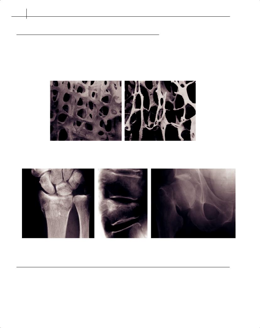
Bone loss leads to thinning, and often perforation, of the trabecular plates (Figure 11.1). Trabecular perforation occurs particularly in situations of increased bone turnover, e.g. after the menopause, and the resulting loss of normal architecture leads to a disproportionate loss of strength for the amount of bone lost. Increased bone turnover is an independent predictor of fracture risk. This may reflect the increase in number of remodelling sites, which can act as a stress riser and increase bone fragility. As a result of accelerated bone loss caused by oestrogen deficiency, postmenopausal osteoporosis initially leads to predominant loss of trabecular bone and frequent trabecular perforation. This typically results in fractures of vertebral bodies and the distal forearm in the sixth and seventh decades of life. In later life, age-related reductions in bone due to remodelling imbalances predominate in both sexes in both cortical and trabecular bone, resulting in the typical manifestation of fracture of the proximal femur.
Epidemiology
The classical osteoporotic fractures are those of the spine, wrist and hip (Figure 11.2) but all fragility fractures in the elderly can be regarded as osteoporotic once pathological fracture (e.g. metastatic disease) has been excluded. Osteoporotic fractures cause considerable morbidity and mortality. Recent estimates suggest that the cost of managing such fractures in the UK is over £1.7 billion a year. One in two women and one in five men are likely to sustain a fracture related to osteoporosis by the age of 90 years. The incidence of osteoporotic fractures is increasing more than expected from the ageing of the population. This may reflect changing patterns of exercise or diet in recent decades.
Classification of osteoporosis
Traditionally, osteoporosis has been classified as primary (includes postmenopausal and age-related bone loss) or secondary (where bone loss is accelerated by the presence of an underlying disease) (Table 11.2). Secondary osteoporosis accounts for up to 40% of cases of osteoporosis in women and 60% of cases in men.
65
66 ABC of Rheumatology
Table 11.1 The World Health Organization’s diagnostic thresholds for bone mineral density at the spine, hip or distal forearm
Diagnosis |
Bone mineral density T score (SD units) |
|
|
Normal |
≥−1 |
Osteopaenia (or low bone mass) |
<−1 but >−2.5 |
Osteoporosis |
≤−2.5 |
Severe osteoporosis |
≤−2.5 plus one or more fragility fractures |
|
|
(a) |
(b) |
Figure 11.1 Comparison of structure of trabecular bone from healthy (a) and osteoporotic (b) subjects, illustrating the architectural damage resulting from trabecular perforation
(a) |
(b), (c) |
Figure 11.2 Typical sites of osteoporotic fracture: wrist (a), vertebrae (b) and hip (c)
Table 11.2 Relatively common causes of secondary osteoporosis
Endocrine |
Gastrointestinal |
Rheumatological |
Malignancy |
Drugs |
|
|
|
|
|
Thyrotoxicosis |
Malabsorption syndrome, e.g. coeliac |
Rheumatoid arthritis |
Multiple myeloma |
Glucocorticoids |
Primary hyper-parathyroidism |
disease, partial gastrectomy |
Ankylosing spondylitis |
Cancer-treatment-induced |
Anticonvulsants |
Cushing’s syndrome |
Inflammatory bowel disease |
|
bone loss (see drugs) |
Heparin |
Hypogonadism, including |
Liver disease, e.g. primary biliary |
|
|
Aromatase inhibitors |
anorexia nervosa |
cirrhosis |
|
|
Androgen-deprivation therapy |
Diabetes type I |
|
|
|
|
|
|
|
|
|