Материал: ABC-of-Rheumatology-ABC-Series-

Osteoarthritis 57
Core |
Options |
|
|
Topical NSAID |
|
|
topical capsaicin |
|
|
oral NSAID |
|
|
opioids |
|
Education/information access |
nutripharmaceuticals |
|
ia steroid |
||
|
||
|
ia hyaluronan |
|
strengthening exercise |
walking aid |
|
aerobic fitness training |
mechanical supports |
|
|
assistive devices |
|
weight loss if overweight/obese |
acupuncture |
|
thermotherapy |
||
reduce adverse mechanics |
||
electrotherapy |
||
|
||
paracetamol |
telephone contact |
|
coping strategies |
||
|
||
|
washout |
|
|
surgery |
Figure 9.10 Summary of the medical management of OA, illustrating the need for a holistic approach. ia = intra-articular
Exercise
Local quadriceps-strengthening exercise can reduce pain and disability and improve the physiological accompaniments of knee OA (muscle weakness, impaired proprioception and balance, tendency to fall). Aerobic activity also reduces pain and disability from OA, improves well-being and sleep quality, and is beneficial for common co-morbidities. Both forms of exercise need to be prescribed. Increased activity and exercise can be accomplished in a variety of ways (e.g. home exercise, group classes), tailored to the patient’s wishes and lifestyle. Pool exercise, wherein people weigh just oneeighth what they weigh on land, can mitigate negative effects of excessive joint loading due to obesity and allow freedom of joint movement and aerobic training for individuals with lower extremity OA.
Reduction of adverse biomechanical factors
Spreading physically hard jobs (e.g. housework, mowing the lawn) at intervals through the day, with breaks in between (“pacing”), can reduce sustained mechanical loading. Weight reduction can improve function and reduce pain in obese and overweight patients and may slow progression of knee and hip OA. Appropriate footwear (thick soft sole, no raised heel, broad forefoot and deep soft uppers) can reduce impact loading in people with knee and hip OA, and wedged insoles can counteract knee varus deformity. Walking sticks and other walking aids reduce loading across OA joints.
Pharmacological treatments
The high prevalence of OA, especially in the elderly, means that co-morbid conditions often exist, and management of OA must take into account these conditions and potential for drug interactions.
Pain is the main reason patients seek help. Long term, the nonpharmacological lifestyle measures mentioned above can all reduce
pain, but drugs are often helpful adjuncts to help quickly reduce pain during exacerbations of symptoms. Paracetamol should be the first oral analgesic to try, based on its excellent safety and reasonable efficacy. Topical non-steroidal anti-inflammatory drugs (NSAIDs) and topical capsaicin are also safe and are particularly useful for hand and knee OA.
Oral NSAIDs, including highly selective COX inhibitors, and weak opioids (e.g. codeine, tramadol) may be considered for those patients who obtain insufficient relief from paracetamol and/or topical agents. The increased risk of gastrointestinal ulceration and bleeding from traditional NSAIDs can be decreased by concomitant prescription of a proton pump inhibitor or misoprostol. The highly selective COX inhibitors, although safer on the gut, may increase the risk of myocardial infarction and stroke, as indeed may many of the traditional NSAIDs. Traditional NSAIDs also cause adverse effects on renal function, especially in the elderly, and have multiple potential drug interactions. Oral NSAIDs and selective COX inhibitors therefore should be given at the lowest effective dose on an as-required, rather than regular, basis. Weak opioids, either alone or in combination with paracetamol, may provide good pain relief, but central nervous system side effects (e.g. constipation, headache, confusion) often limit their usefulness.
Nutraceuticals provide an alternative in older, high-risk patients with co-morbidity because they have no associated renal or gastrointestinal side effects and are very popular with patients. Glucosamine is contraindicated in patients with shellfish allergy. The initiation and use of either glucosamine or chondroitin sulphate requires monitoring of glucose in diabetics, as these agents are associated with mild insulin resistance in animals.
Intra-articular corticosteroid injection is a valuable treatment that often gives quick effective relief of pain that may last just a few weeks to a few months. It is particularly useful to tide a patient over an important event (e.g. family wedding, holiday) and to improve pain during initiation of other interventions such as an exercise programme. A variety of hyaluronan preparations are also available, given as a single injection or a course of one per week for 3–5 weeks. Although a modest, relatively prolonged (several months) improvement in pain may result, the cost and logistics of this treatment are limiting.
Surgical
The success of prosthetic joint replacements has greatly advanced management of end-stage hip and knee OA. Surgery is also used increasingly now at the shoulder, elbow, and thumb base. Although issues of funding, waiting times, choice of prosthesis and revision have to be faced, there is no doubt that such surgery can transform a patient’s life. Other surgical approaches (osteotomy, arthrodesis or joint fusion) may also be useful in specific circumstances. Arthroscopic debridement and lavage is indicated if a patient with OA describes mechanical locking. Symptomatic improvement following joint lavage alone can last several months.
The criteria for referral for consideration of joint replacement are not universally agreed upon, but include uncontrolled pain and severe impairment of function despite conservative treatment. Age,

58 ABC of Rheumatology
in itself, is not a contraindication. Autologous chondrocyte transplantation is a procedure that is currently typically reserved for young patients with severe chondral defects.
In summary, OA is a condition of increasing prevalence characterized by a phasic progression with periods of relative quiescence and flare. An individualized and holistic approach to management is essential as the best means for relieving pain, minimizing disability and improving quality of life.
eds. Osteoarthritis: diagnosis and medical/surgical management, 4th edn. Lippincott Williams and Wilkins, Philadelphia, PA, 2007: 27–49.
Roos EM. Joint injury causes knee osteoarthritis in young adults. Current Opinion in Rheumatology 2005; 17: 195–200.
van Saase JL, van Romunde LK, Cats A, Vandenbroucke JP, Valkenburg HA. Epidemiology of osteoarthritis: Zoetermeer survey. Comparison of radiological osteoarthritis in a Dutch population with that in 10 other populations. Annals of the Rheumatic Diseases 1989; 48: 271–280.
References
Arden N, Nevitt M. Osteoarthritis: epidemiology. Best Practice and Research Clinical Rheumatology 2006; 20: 3–25.
CDC (Centers for Disease Control and Prevention). Morbidity and Mortality Weekly Report, vol. 50, no. 7. US Department of Health and Human Services, CDC, Atlanta, Georgia. February 23, 2001: 123.
CDC (Centers for Disease Control and Prevention). Behavioral Risk Factor Surveillance System Survey Data. US Department of Health and Human Services, CDC, Atlanta, Georgia, 2005.
CDC (Centers for Disease Control and Prevention). Morbidity and Mortality Weekly Report, vol. 55, no. 40. US Department of Health and Human Services, CDC, Atlanta, Georgia. October 13, 2006: 1091.
CDC (Centers for Disease Control and Prevention). Morbidity and Mortality Weekly Report, vol. 56, no. 17. US Department of Health and Human Services, CDC, Atlanta, Georgia. May 4, 2007: 424.
Englund M, Paradowski PT, Lohmander LS. Association of radiographic hand osteoarthritis with radiographic knee osteoarthritis after meniscectomy. Arthritis and Rheumatism 2004; 50: 469–475.
Engstrom-Laurent A, Hallgren R. Circulating hyaluronic acid levels vary with physical activity in healthy subjects and in rheumatoid arthritis patients. Relationship to synovitis mass and morning stiffness. Arthritis and Rheumatism 1987; 30: 1333–1338.
Pincus T, Mitchell JM, Burkhauser RV. Substantial work disability and earnings losses in individuals less than age 65 with osteoarthritis: comparisons with rheumatoid arthritis. Journal of Clinical Epidemiology 1989; 42: 449–457.
Poole AR, Guilak F, Abramson SB. Etiopathogenesis of osteoarthritis. In: Moskowitz RW, Altman RD, Hochberg MC, Buckwalter JA, Goldberg VM,
Further reading
American College of Rheumatology. Recommendations for the medical management of osteoarthritis of the hip and knee. Arthritis and Rheumatism 2000; 43: 1905–1915.
Arden N, Nevitt MC. Osteoarthritis: epidemiology. Best Practice and Research. Clinical Rheumatology 2006; 20: 3–25.
Brandy KD, Doherty M, Lohmander LS, eds. Osteoarthritis, 2th edn. Oxford University Press, Oxford, UK, 2003.
Felson DT, Lawrence RC, Dieppe PA et al. Osteoarthritis: new insights. Part 1: the disease and its risk factors. Annals of Internal Medicine 2000; 133: 635–646.
Jordan KM, Arden N, Doherty M et al. EULAR recommendations: an evidence based medicine approach to the management of knee osteoarthritis.
Annals of the Rheumatic Diseases 2003; 62: 1145–1155.
Moskowitz RW, Altman RD, Hochberg MC, Buckwalter JA, Goldberg VM, eds. Osteoarthritis: diagnosis and medical/surgical management, 4th edn. Lippincott Williams and Wilkins, Philadelphia, PA, 2007.
Zhang W, Doherty M, Arden N et al. EULAR recommendations: an evidencebased medicine approach to the management of hip osteoarthritis. Annals of the Rheumatic Diseases 2005; 64: 669–681.
Zhang W, Doherty M, Leeb BF et al. EULAR evidence-based recommendations for the management of hand osteoarthritis. Annals of the Rheumatic Diseases 2007; 66: 377–388.
Acknowledgements
We would like to express sincere appreciation to Shelby Addison for her invaluable assistance with graphics and permissions for figures.

CHAPTER 10
Gout, Hyperuricaemia and
Crystal Arthritis
Martin Underwood1 and Ade Adebajo2
1University of Warwick, Coventry, UK
2University of Sheffield, Sheffield, UK
OVERVIEW
•Asymptomatic hyperuricaemia does not need treating.
•Steroids, non-steroidal anti-inflammatory drugs (NSAIDs), or colchicine can be used to treat acute gout; oral steroids might provide the best balance of risks and benefits.
•Patients with recurrent gout twice or more a year should be offered urea-lowering medication.
•Target serum urate for patients on urate-lowering drugs is <0.30 or <0.36 mmol/l.
•Dose of urate-lowering medication should be titrated according to response; many patients with recurrent gout get inadequate doses of urate-lowering drugs.
Gout and hyperuricaemia
Gout is a common metabolic disorder, typically presenting as an acute monoarthritis, most commonly of the first metatarsal phalangeal joint. The term “gout” includes an acute attack, the propensity for repeated episodes and also for chronic gouty arthritis. The underlying problem is a build-up of urate, a purine breakdown product. Humans, and some primates, lack uricase, which in other mammals oxidizes urate to allantoin, which is readily soluble. Both an increased dietary purine intake and an increased breakdown of endogenous proteins (e.g. cancer treatment or haematological malignancy) can increase urate levels.
Urate excretion is mainly renal. The rate of renal excretion is affected by urine flow, pH and competition for renal tubular exchange (e.g. diuretics). Patients whose problems are primarily due to increased purine turnover will have a high urinary urate, and those whose problems are primarily renal will have a low urinary urate. This distinction is rarely of clinical importance. For uric acid crystals to form, the serum needs to be saturated with urate, i.e. >0.42 mmol/l (>7.0 g/dl). This is, coincidentally, the upper limit of the normal range in men and postmenopausal women in many laboratories. For premenopausal women, the upper limit of the normal range for serum urate is 0.36 mmol/l (>6.0 mg/dl).
ABC of Rheumatology, 4th edn. Edited by Ade Adebajo. ©2010 Blackwell Publishing Ltd. 9781405170680.
Epidemiology
Prevalence is probably around 1% in white populations. Gout is rare in premenopausal women. However, there are few reliable data on how many people are affected each year, or how many people are taking prophylactic drugs. As gout is associated with increasing age and obesity, it is likely to become commoner. Some non-white populations are more prone to gout/hyperuricaemia; e.g. the prevalence of gout is 6.4% and 3.6%, respectively, in New Zealanders of Maori and European origin. There is generally a higher prevalence of gout in indigenous ethnic groups around the Pacific Rim. In Malaysia, the three largest ethnic groups are Malay, Chinese and Tamil. All have higher mean levels of uric acid in serum than most white populations. Environmental factors also play a part; for example, changes to from a traditional island lifestyle to a more Westernized diet increased prevalence of gout 9-fold, to Maori levels, in Tokelauan islanders who migrated to New Zealand. The prevalence of gout may also be higher in black Afro-Caribbean ethnic groups. In sub-Saharan Africa gout is particularly associated with high alcohol intake in all socio-economic groups. A particular problem for some groups in sub-Saharan Africa is saturnine gout due to the effects of lead, absorbed from containers used for homemade alcoholic drinks, on renal tubular function.
Risk factors
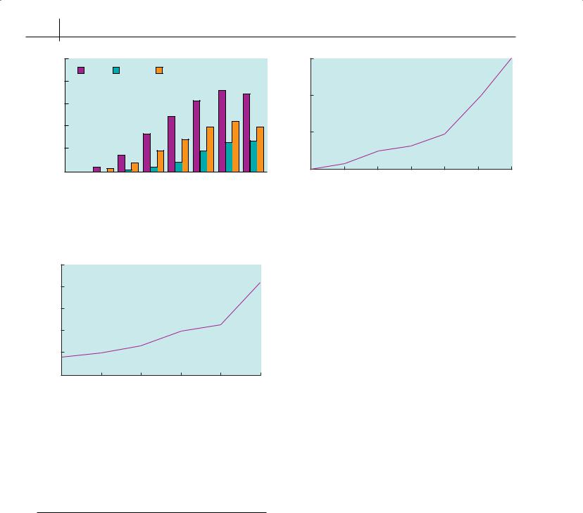
Age and sex—Gout becomes commoner with increasing age (Figure 10.1). In men the reported prevalence ranges from <0.5% in those aged under 35 to over 7% in those aged over 75. It is rare in premenopausal women but increases to 2.5–3.0% in those aged over 75. The later age of onset in women may relate to the uricosuric effects of oestrogens.
Obesity—Relative risk of gout increases with increasing body mass index (BMI). Compared to people with a BMI of 21–25, those with BMI of >35 are four times as likely to develop gout (Figure 10.2).
Diet—Each additional daily portion of meat per day increases the risk of gout by 20%. Purine-rich vegetables do not appear to increase the risk of gout, while consuming more dairy products reduces the risk of developing the disease (Table 10.1).
59
60 ABC of Rheumatology
|
100 |
|
|
|
|
|
|
|
patients) |
|
Men |
Women |
|
Total |
|
|
|
80 |
|
|
|
|
|
|
|
|
1000(per |
|
|
|
|
|
|
|
|
60 |
|
|
|
|
|
|
|
|
|
|
|
|
|
|
|
|
|
Prevalence |
40 |
|
|
|
|
|
|
|
20 |
|
|
|
|
|
|
|
|
|
|
|
|
|
|
|
|
|
|
0 |
25–34 |
35–44 |
45–54 |
55–64 |
65–74 |
75–84 |
≥85 |
|
<24 |
|||||||
|
|
|
Age group (years) |
|
|
|
||
Figure 10.1 Prevalence of gout; data from general practice research database. Reproduced with permission from Underwood (2006)
|
2.5 |
|
|
|
|
|
|
gout |
2.0 |
|
|
|
|
|
|
risk of |
|
|
|
|
|
|
|
|
|
|
|
|
|
|
|
Relative |
1.5 |
|
|
|
|
|
|
|
|
|
|
|
|
|
|
|
1.0 |
|
|
|
|
|
>50 |
|
0 |
0.1–4.9 |
5.0–9.9 |
10–14.9 |
15.0–29.9 |
30.0–49.9 |
Alcohol (g/day)
Figure 10.3 Effect of total alcohol intake on the relative risk of a first attack of gout; data from the health professionals follow-up study. Reproduced with permission from Underwood (2006)
|
5 |
|
|
|
|
|
of gout |
4 |
|
|
|
|
|
3 |
|
|
|
|
|
|
risk |
|
|
|
|
|
|
Relative |
2 |
|
|
|
|
|
1 |
|
|
|
|
|
|
|
|
|
|
|
|
|
|
0 |
|
|
|
|
>35.0 |
|
<21.0 |
21.0–22.9 |
23.0–24.9 |
25.0–29.9 |
30.0–34.9 |
Body mass index
Figure 10.2 Obesity and the incidence of a first attack of gout in men; data from the health professionals follow-up study. Reproduced with permission from Underwood (2006)
Table 10.1 Effect of diet and alcohol on incidence of a first attack of gout in men; data from the health professionals follow-up study. Reproduced with permission from Underwood (2006)
Portion |
Relative risk (95% CI) |
|
|
Alcohol |
|
Beer 335 ml |
1.49 (1.32 to 1.70) |
Sprits 44 ml |
1.15 (1.04 to 1.20) |
Wine 118 ml |
1.04 (0.88 to 1.22) |
Food: |
|
Meat |
1.21 (1.04 to 1.41) |
Seafood (fish)* |
1.07 (1.01 to 1.12) |
Purine rich vegetables |
0.97 (0.79 to 1.19) |
Total daily products |
0.82 (0.75 to 0.90) |
Low-fat daily products |
0.79 (0.71 to 0.87) |
High-fat daily products |
0.99 (0.89 to 1.10) |
|
|
Data from health professionals follow-up study. *Calculated from weekly intake.
Dietary fructose may also increase the risk of developing gout. This occurs naturally and is also present in high-fructose corn syrup (HFCS), which is commonly used as a sweetener for soft drinks and other foods in the USA. The use of HFCS is uncommon outside the USA.
Drugs—A number of drugs can increase serum urate; this is most commonly due to diuretics. Aspirin and salicylates at low doses decrease urate excretion, but at high doses (4–6 g/day) they have a uricosuric effect.
Alcohol—Compared to non-drinkers, people consuming >50 g alcohol per day are 2.5 times as likely to develop gout (Figure 10.3). While there is a strong relationship between beer intake and gout, there is only a weak relationship between intake of spirits and gout. There does not appear to be a relationship between wine intake and gout. Alcohol is catabolized to ketones that compete with urate for excretion by the renal tubule. Beer typically contains substantial amounts of purines, from yeast, which are catabolized to urate by gut bacteria. Alcohol may also increase the dose of allopurinol needed by decreasing the conversion of allopurinol to its effective metabolite, oxipurinol.
Relationship between gout and hyperuricaemia
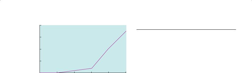
Hyperuricaemia is necessary for the development of gout. Crystal deposition can only occur when the serum is saturated with urate: ≥0.42 mmol/l. This may be different from some laboratories’ normal ranges, which are based on population norms. Only a minority of people with hyperuricaemia develop gout. For example, the annual incidence of gout is only 6% in people with a urate of 0.60 mmol/l (Figure 10.4). Serum urate can fall during an acute
Gout, Hyperuricaemia and Crystal Arthritis |
61 |
|
|
|
|
|
8 |
|
|
|
|
|
(%) |
|
|
|
|
|
|
of gout |
6 |
|
|
|
|
|
|
|
|
|
|
|
|
instance |
4 |
|
|
|
|
|
|
|
|
|
|
|
|
Annual |
2 |
|
|
|
|
|
|
|
|
|
|
|
|
|
0 |
|
|
|
|
≥0.6 |
|
<0.36 |
0.36–0.41 |
0.42–0.47 |
0.48–0.53 |
0.54–0.59 |
Serum urate concentration (mmol/l)
Figure 10.4 Relationship between serum urate and the annual incidence of gout; data from normative ageing study. Reproduced with permission from Underwood (2006)
attack, and patients on urate-lowering medication can still be affected until crystal deposits have cleared from the joints. Thus, demonstrating a raised serum urate is not an essential prerequisite for diagnosing gout.
Hyperuricaemia and cardiovascular disease
There is a well-recognized association between hyperuricaemia and cardiovascular disease. It is not clear whether hyperuricaemia is an independent risk factor for cardiovascular disease. Thus, screening for hyperuricaemia in those with a high cardiovascular risk is not indicated. However, assessing cardiovascular risk in people presenting with gout is worthwhile.
Clinical features
Acute gout
Typically gout presents as a rapid onset of a monoarthritis associated with severe pain and inflammation classically affecting the first metatarsophalangeal joint (podagra) Low-grade fever, general malaise and anorexia may accompany the joint symptoms. Onset may follow a drinking bout, or local trauma. Untreated, acute gout usually resolves spontaneously within 7–10 days but can on occasion last several weeks. The other most commonly affected areas are the other joints in the foot, ankle, knee, wrist, finger and elbow. The affected joint is warm, tender and swollen, and in most cases, the overlying skin is erythematous. The predilection for peripheral joints is probably because crystals are more likely to form in cooler joints. Typically, the attack occurs during the night. After the acute attack patients may be symptom-free for months or years.
Diagnosis—The diagnosis of acute gout is usually clinical. The European League Against Rheumatism (EULAR) recommendations for gout suggest that:
‘the rapid development of severe pain, swelling and tenderness that reaches its maximum within just 6–12 hours, especially with overlying erythema, is highly suggestive of crystal inflammation though not specific for gout’
Table 10.2 Investigation of patients with gout
Diagnosis |
Causes |
Co-morbidities |
|
|
|
|
|
Serum urate |
Full blood count |
Blood pressure |
|
May fall during an |
To exclude myeloand |
Cholesterol |
|
attack |
lymphoproliferative |
||
|
|||
Joint aspiration |
disorders, secondary |
Blood sugar |
|
polycythaemia, haemolytic |
|
||
Consider if diagnosis |
Thyroid function |
||
anaemia, haemoglobinopa- |
|||
uncertain; will enable |
15% of patients |
||
thies; white count may be |
|||
the alternative |
with gout have |
||
slightly raised; if very high |
|||
diagnosis of pseudog- |
hypothyroidism |
||
consider septic arthritis |
|||
out to be made if |
|
||
|
Uric acid |
||
pyrophosphate crystals |
Liver enzymes |
||
excretion |
|||
are present; note, |
Possible alcohol abuse |
||
Consider if strong |
|||
however, that both |
Renal function |
||
family history of |
|||
types of crystals can |
Drug doses might need |
||
gout, if onset |
|||
coexist |
adjusting if renal function is |
||
under age 25 or if |
|||
|
poor |
||
|
renal stones |
||
|
|
||
|
Review medication |
present |
|
|
Diuretics and some other |
|
|
|
drugs increase urate |
|
|
|
Investigations if |
|
|
|
pseudogout suspected |
|
|
|
Calcium, magnesium, |
|
|
|
ferritin, thyroid function |
|
|
|
|
|
and that:
‘for typical presentations of gout (such as podagra with hyperuricaemia) a clinical diagnosis alone is reasonably accurate but not definitive without crystal confirmation.’
The gold standard is demonstrating urate crystals in synovial fluid. However, few generalists (or their patients) will relish aspirating an acutely inflamed first metatarsophalangeal joint. Urate crystals are strongly negatively birefringent on polarizing microscopy. Pyrophosphate crystals, which are weakly positively birefringent under polarized light, are found in pseudogout. Both types of crystals coexist in about 10% of crystal-associated synovial effusions. In the acute situation the important differential diagnosis is septic arthritis. If septic arthritis is suspected, then urgent specialist assessment is needed. Bursitis of the first metatarsophalangeal joint can mimic podagra and is often mislabelled and mistreated as gout, especially in young women.
Investigations—All patients with a suspected first episode of gout should be investigated to obtain some confirmatory evidence to support the diagnosis, to look for underlying causes and to identify associated co-morbidities (Table 10.2). X-rays are not helpful in the diagnosis of acute gout.
Chronic gout
Chronic, polyor oligoarticular gout can cause inflammatory arthritis in older people, especially those on diuretics. No diagnostic pattern exists, although lower-limb-joint involvement is