Материал: ABC-of-Rheumatology-ABC-Series-

Treatment of Rheumatoid Arthritis |
77 |
|
|
|
|
Box 13.1 Side effects of methotrexate
•Mucosal ulceration
•Alopecia
•Gastrointestinal—nausea and vomiting, oesophagitis, anorexia, diarrhoea, gingivitis, GI bleeding, GI perforation, pancreatitis, elevated transaminases, cirrhosis
•Bone marrow suppression—anaemia, leucopenia, thrombocytopenia, aplastic anaemia, malignancy—lymphoproliferative disorders
•Infections
•Interstitial pneumonitis
•Renal impairment
•Teratogenesis
Disease-modifying antirheumatic drugs
Gold—Originally intended for the treatment of infectious diseases, gold is one of the oldest of the disease-modifying antirheumatic drugs (DMARDs) and has been in use for almost a century for the treatment of RA. An intramuscular drug of proven efficacy, radiological improvement with decrease in radiological damage bore evidence of its disease-modifying capacity. However, weekly injections may be cumbersome, and an oral form proved inefficacious. This, together with the fact that over half of drug discontinuations were reported to be the result of toxicity (such as severe skin rash and nephrotoxicity), heralded a decline in its popularity over the years.
Methotrexate—A dihydrofolate reductase inhibitor originally used for its anti-proliferative effects in the treatment of cancer, methotrexate is now an anchor drug among DMARDs and a gold standard against which all emerging therapies are compared. An oral drug administered on a weekly basis, its anti-inflammatory mechanisms of action are thought to differ from its anti-malignant effects, and are largely related to its induction of adenosine release to the inflammatory environment. Compared with gold it has an excellent side-effect profile, the only frequent problem being post-dos- age nausea, which frequently responds to folic acid (Box 13.1).
Sulfasalazine—Sulfasalazine, the first drug developed specifically for the treatment of RA, was first synthesized in the 1940s. It is composed of sulfapyridine and 5-aminosalicylic acid moieties and should be avoided in patients allergic to sulfa medications. Plasma half-life is greatly influenced by acetylation status, and slow acetylators are more likely to develop serious toxicities. While minor upper GI side effects and rashes are common, drug-induced hepatitis, cytopenias and Stevens–Johnson syndrome may also occur.
Hydroxychloroquine—Hydroxychloroquine is an antimalarial with proven efficacy in the treatment of RA, particularly in early and mild disease. Unlike its sister drug, chloroquine, occurrence of retinopathy is extremely rare. However, evidence for radiological protection has been unconvincing, and this benign medication in most often used in conjunction with other DMARDs in combination therapy rather than alone.
Box 13.2 Other DMARDs in use
•Penicillamine
•Azathioprine
•Cyclophosphamide
•Ciclosporin
•Tacrolimus
•Mycophenolate mofetil
•Minocycline
Leflunomide—The youngest member among the DMARDs, leflunomide inhibits the de novo synthesis of pyrimidines by inhibiting dihydroorotate dehydrogenase, and this action principally affects lymphocytes which lack salvage pathways for pyrimidine synthesis. It may be useful in patients who have failed to respond to methotrexate, but can also be administered together with methotrexate to improve response. It has a long half-life, requiring a loading dose for 1–3 days. As a long washout period of up to 2 years is suggested prior to conception, careful planning is needed in premenopausal women.
Some of the other DMARDs in use are listed in Box 13.2.
Combination therapy
Although DMARDs represent a marked improvement over previous symptom-oriented therapies, response to monotherapy is often partial at best, and discontinuation, whether due to toxicity or lack of response, is commonplace. It has been suggested that using these medications with different but complementary mechanisms of action in combination not only allows for greater efficacy, but also limits effective required dosage and hence toxicity. Various combinations, including step-up and step-down regimens have been tried, often with the inclusion of methotrexate.
Biological-response modifiers
A major development in the treatment of RA in the last decade was the emergence of biological-response-modifying therapy. Previous attempts at drug development have largely been empirical efforts. Understanding of the molecular and cellular mechanisms that contribute to the generation and maintenance of the inflammatory processes that culminate in synovial inflammation and joint destruction has escalated astronomically in recent decades. These fundamental elements of the inflammatory cascade, whether it be a cytokine or an inflammatory cell subset, have become the targets of new treatment modalities. These drugs are administered parenterally, and the onset of action, unlike DMARDs, is rapid.
Tumour necrosis factor antagonists
Tumour necrosis factor (TNF) is a pivotal cytokine released in excess in RA, and is a major contributor to synovial inflammation and cartilage destruction. Blockade of its actions by the human TNF receptor 2–immunoglobulin constant region fusion protein, etanercept, resulted in the first success of biological-

78 ABC of Rheumatology
response-modifying therapy in RA. Since then, monoclonal antibodies to human TNF have come into use, whether chimeric (infliximab) or fully humanized (adalimumab). These agents have been demonstrated to be efficacious in the treatment of RA on clinical, radiological and laboratory measures, particularly when used in combination with methotrexate.
Anakinra
The next cytokine to be targeted for therapeutic use was inter- leukin-1 (IL-1) and anakinra is a recombinant human IL-1 receptor antagonist. Its short half-life means that subcutaneous injections have to be given on a daily basis. Used alone or in combination with methotrexate, anakinra produces significant if modest clinical improvement in RA. Radiological improvement, including rates of progression of joint-space narrowing and erosion, have been more striking, however.
Rituximab
The B-lymphocyte is not only the source of inflammatory cytokines and antibodies important to the pathogenesis of the disease such as rheumatoid factor and anti-cyclic citrullinated peptide; B-cell help is a vital contributor to T-cell activation and antigen presentation. It is therefore little surprise that the B-lymphocyte may be a suitable target for RA therapy, despite previous dogma that RA is predominantly a T-cell-mediated disease. Rituximab targets the B-cell surface marker, CD20, which is expressed from the pre-B- cell stage through to the mature memory B-cell. Binding of this chimeric monoclonal antibody depletes CD20+ B-cells in a transient manner. Rituximab, whether alone or in combination with methotrexate, is effective at suppressing inflammatory parameters and limiting structural joint damage in RA, although seronegative patients have responded less well. While the risk of infection remains a concern with B-cell depletion, this has not been a problem based on available clinical trial data. However, clinicians should be alerted to reports of rare neurological diseases, such as progressive multifocal leukoencephalopathy, caused by infection with the JC polyoma virus.
Abatacept
T-lymphocyte activation and proliferation requires a dual stimulatory signal that involves both the T-cell and the antigen-presenting cell. Interruption of any individual part of this signalling complex, such as CTLA-4, the target of abatacept, disrupts T-cell contributions to the inflammatory environment. Abatacept has proven benefits in slowing disease activity and joint damage in RA, including in patients who have failed to respond to anti-TNF therapy, although it has not been recommended for use in Britain in a
preliminary judgment by the National Institute for Health and Clinical Excellence.
Identification of coexisting problems
RA has long been regarded as an indolent disease, until recently when it has been recognized that RA may be associated with increased mortality. Life expectancy may be shortened by as much as 7 years in men and 3 years in women. Yet few have been able to attribute deaths directly to RA itself. Clinicians should therefore be constantly alerted to the development of other co-morbidities in RA patients, of which cardiovascular disease is the most important. Occurrence of cardiovascular disease has been reported in up to 42% of RA patients, and scrupulous management and correction of cardiovascular risk factors is of utmost importance to disease outlook. Although vasculitis, secondary amyloidosis and lymphoproliferative malignancies have been associated with the disease itself, these are rare, and renal, pulmonary, GI and infectious diseases are much more common. Furthermore, antirheumatic pharmacotherapy itself may compound these problems. The presence of co-morbidities is a known predictor for mortality in RA patients, and due attention must be given to its early identification.
Complimentary therapy
Although our discussion has focused on the pharmacotherapy of RA, it should be remembered that one of the main goals of management is restoration of function. In this respect, the roles of physiotherapy and occupational therapy and meticulous foot care cannot be overlooked. Advances in orthopaedic surgery have also benefited situations such as atlanto-axial subluxation, arthroplasties and tendon transfer and repair surgeries.
Further reading
Blom M, van Riel PL. Management of established rheumatoid arthritis with an emphasis on pharmacotherapy. Best Practice & Research. Clinical Rheumatology 2007; 21: 43–57.
Chan ESL, Cronstein BN. Drugs that modulate the immune response. In: Samter’s Immunologic Diseases, 6th edn. Lippincott Williams & Wilkins 2001: 1213–1223.
Fleischmann RM. Comparison of the efficacy of biologic therapy for rheumatoid arthritis: can the clinical trials be accurately compared? Rheumatic Diseases Clinics of North America 2006; 32 (Suppl. 1): 21–28.
Goldblatt F, Isenberg DA. New therapies for rheumatoid arthritis. Clinical and Experimental Immunology 2005; 140: 195–204.
Lee SJ, Kavanaugh A. Pharmacological treatment of established rheumatoid arthritis. Best Practice & Research. Clinical Rheumatology 2003; 17: 811–829.

CHAPTER 14
Spondyloarthritides
Andrew Keat1 and Robert Inman2
1Northwick Park Hospital, Harrow, UK
2Toronto Hospital Western Division, Toronto, Canada
OVERVIEW
•Spondyloarthritides as a group occur with a similar prevalence to rheumatoid arthritis.
•The various spondyloarthritic syndromes share common clinical lesions, especially enthesitis, oligoarthritis, sacroiliitis, iritis, psoriasiform skin and mucosal lesions and overt or covert inflammatory bowel disease.
•Inheritance of HLA-B27 and other genes is common to all spondyloarthritides, the prevalence of these disorders varying with the local prevalence of HLA-B27.
•Diagnosis of ankylosing spondylitis is often long delayed; identification of inflammatory back pain is a key determinant in making the diagnosis early.
•Use of anti-tumour necrosis factor biologic drugs has revolutionized the treatment of severe ankylosing spondylitis.
The spondyloarthritides (SpA) comprise a group of syndromes that are distinct from rheumatoid arthritis and are characterized by inflammation of the spine in many, but not all, cases. Other key features include asymmetric oligoarthritis, enthesitis, psoriatic skin and mucous membrane lesions, and eye and bowel inflammation. Tests for rheumatoid factor, anti-cyclic citrullinated peptide antibody and other autoantibodies are negative, but there is a strong association with the human leukocyte antigen (HLA) B27. Spondyloarthritides occur in both adults and children, although spinal involvement is rare in children. A working definition has been provided by the European Spondyloarthritis Study Group (Box 14.1). A diagnosis of SpA requires one or two of the entry criteria plus one other. Skin, eye and bowel disease may become apparent only with the passage of time.
The classical forms of spondyloarthritis (also called “spondyloarthropathies”) and the key physical features are listed in Table 14.1.
ABC of Rheumatology, 4th edn. Edited by Ade Adebajo. ©2010 Blackwell Publishing Ltd. 9781405170680.
Box 14.1 The European Spondyloarthropathy Study Group
Criteria for Spondyloarthropathy
• Inflammatory spinal pain (defined as low back pain with morning stiffness, better on exercise, in patients <40 years old)
or
•Asymmetric or predominantly lower limb synovitis plus
•Any one or more of the following: psoriasis, inflammatory bowel disease, alternating buttock pain, enthesopathy, sacroiliitis
Together, spondylarthritides are roughly as common as rheumatoid arthritis in Europe and North America, although their prevalence varies in other areas, generally reflecting the prevalence of HLA-B27 in that population. Their prevalence and that of associated conditions is presented in Table 14.2.
Ankylosing spondylitis
Ankylosing spondylitis (AS) is an aseptic inflammatory condition of the joints and entheses of the spine. AS occurs in 0.2% of the general population, in 2% of the B27-positive population and in 20% of B27-positive individuals with an affected family member. Males predominate with a male:female ratio ranging from 2.5:1 to 5:1. AS typically begins in young adulthood, but symptoms may arise in adolescence or earlier. Up to 15% of children with juvenile idiopathic arthritis are classified as having juvenile-onset spondyloarthritis. Such children present with pauciarticular peripheral arthritis with a predilection for the tarsal joints; axial complaints, with the development of radiographic sacroiliitis, tend only to develop in late teenage years or later.
The first symptom of AS is usually inflammatory back pain—the insidious onset of low back pain and/or buttock pain that persists for more than 3 months, awakens the patient from sleep, is accompanied by early morning stiffness and is typically improved by exercise. Fatigue often accompanies inflammatory back pain, although it may also be present in fibromyalgia and other conditions. Inadequately controlled inflammation leads to persistent stiffness and progressive loss of spinal mobility.
79

80 ABC of Rheumatology
Table 14.1 Examples of spondyloarthropathies
Syndromes |
Features |
|
|
Ankylosing spondylitis |
Sacroiilitis |
|
Enthesitis |
|
Spondylitis |
Psoriatic arthritis |
Oligoarthritis |
|
Dactylitis |
|
Skin and mucous membrane |
|
inflammation |
|
Psoriasis |
Reactive arthritis (Reiter’s syndrome) |
Genito-urinary inflammation, iritis |
Enteropathic arthritis |
Small and large bowel inflammation |
Undifferentiated spondyloarthritis |
Possible infectious trigger |
Childhood spondyloarthritis |
Associated with HLA-B27 |
|
|
Box 14.2 The modified New York criteria for ankylosing spondylitis (1984)
A. Diagnosis
1. Clinical criteria
a. Low back pain and stiffness >3 months with improvement on exercise, not relieved by rest
b. Limitation of spinal motion in both sagittal and frontal planes
c. Limitation of chest expansion 2. Radiologic criteria
Sacroiliitis: Grade >2 bilaterally or Grade 3–4 unilaterally B. Grading
1. Definite ankylosing spondylitis if the radiologic criterion is associated with >1 clinical criterion
2. Probable ankylosing spondylitis if:
a. the three clinical criteria are present
b. the radiologic criterion is present without any signs or symptoms satisfying the clinical criteria
Table 14.2 Prevalence of spondyloarthropathies
|
Prevalence (%) per |
male:female |
|
100,000 |
|
|
|
|
Ankylosing spondylitis |
0.2 |
3.5 |
Psoriasis |
2000 |
1.0 |
Psoriatic arthritis |
20–100 |
1.3 |
Reactive arthritis |
16 |
3.0 |
Crohn’s disease |
30–75 |
1.0 |
Ulcerative disease |
50–100 |
0.8 |
Enteropathic arthritis |
1–20% of inflammatory |
* |
|
bowel disease |
|
|
|
|
*Peripheral arthritis occurs more in women; sacroiliitis and spondylitis more often affect men
Box 14.3 Characteristics of inflammatory back pain (in patients <50 years old)
•Morning stiffness >30 minutes duration
•Improvement in back pain with exercise but not with rest
•Awakening because of back pain during the second half of the night only
•Alternating buttock pain
If none of four parameters present post-test probability of AS 1.3%
If one of four parameters present post-test probability of AS 2.6%
If two of four parameters present post-test probability of AS 10.8%
If three or more parameters present post-test probability of AS 39.4%, with sensitivity of 33.6% and specificity of 97.3%
(See Rudwaleit et al.)
The diagnosis is based on the modified New York criteria (Box 14.2). Radiographic assessment is a key element of these criteria: classical changes in the sacroiliac joints include erosions in the joint line, pseudowidening, subchondral sclerosis and finally ankylosis, reflected as obliteration of the sacroiliac joint. Radiographs of the spine may reveal squaring and “shiny corners” of the vertebral bodies and, later, syndesmophytes and facet-joint fusion (Figure 14.1). As radiographic sacroiliitis often develops late, early diagnosis may be based on symptoms of inflammatory back pain (Box 14.3) combined with magnetic resonance imaging (MRI) evidence of sacroiliitis (Figure 14.2).
HLA-B27 is rarely the definitive factor for diagnosis, but when the clinical suspicion is high, the test has reasonably high sensitivity and specificity.
Up to 30% of patients with AS also develop peripheral arthritis. Typically this is asymmetrical oligoarthritis affecting leg joints,
most commonly the knee. Involvement of the hip can occur at any point in the course of AS and may be highly destructive. Enthesitis—inflammation at attachments of tendon or ligament to bone—is also a characteristic feature of AS. Enthesitis at the calcaneal attachments of the Achilles tendon, usually accompanied by Achilles tendon bursitis (Figure 14.3) and plantar fascia, producing sometimes disabling heel pain, is highly characteristic of AS, although it also occurs in other SpAs. Dactylitis, usually affecting a toe (“sausage toe”) (Figure 14.4), is also strongly suggestive of an SpA.
Ocular inflammation, usually acute anterior uveitis (iritis), occurs at some time in up to 40% of AS patients. Acute anterior uveitis typically causes pain, photophobia and, if untreated, impairment in visual acuity. Typically, it is unilateral and recurrent. Uncommon extra-articular manifestations of AS include aortic insufficiency, cardiac conduction defects and pulmonary fibrosis.
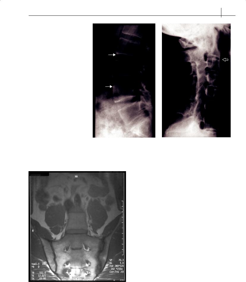
Spondyloarthritides 81
*
Figure 14.1 Radiographs of the spine showing early changes of “shiny corners” ( ) late changes of syndesmophytes ( ) and facet-joint fusion ( )
Assessment of ankylosing spondylitis
In recent years several instruments have been devised for measuring disease activity, overall function, severity and progression of AS. Those most widely used are detailed below.
Disease Activity—Bath Ankylosing Spondylitis Disease Activity Index (BASDAI). A patient-completed set of six visual analogue scales assessing symptoms. The erythrocyte sedimentation rate and C-reactive protein are typically elevated, but levels do not usefully indicate inflammatory activity of spinal disease.
Overall patient function—Bath Ankylosing Spondylitis Functional Index (BASFI). A similar patient-completed set of 10 visual analogue scales assessing normal daily activities.
Spinal mobility—Bath Ankylosing Spondylitis Metrology Index (BASMI). A composite score derived from measurements of spinal mobility.
|
Radiologic progression—Modified Stoke AS Spinal Score (mSASSS). |
|
This uses lateral radiographs of the cervical and lumbosacral spine |
|
and can detect change over 2 years. It evaluates the anterior part of |
|
the lumbar spine and cervical spine and assesses chronic changes |
|
at each level with a score of 0 to 3 (0 = normal; 1 = erosion, sclerosis |
Figure 14.2 MRI scan showing right-sided sacroiliitis |
or squaring; 2 = syndesmophyte; 3 = bridging syndesmophyte). |