Материал: ABC-of-Rheumatology-ABC-Series-
102 ABC of Rheumatology
oligoarticular and follows bacterial infection in the gut (Salmonella, Shigella, Campylobacter, Yersinia), although in the older child and adolescent it is important to consider sexually acquired infection (Chlamydia, gonorrhoea). Rheumatic fever (a form of reactive arthritis that follows pharyngeal streptococcal infection) is uncommon in the UK, but common in developing countries. Lyme disease following tick-transmitted infection with Borrelia burgdorferi is suggested by the presence of an oligoarthritis, erythema chronicum migrans, and a travel history to an endemic area.
Chronic arthritis (JIA)
In the absence of sepsis or trauma, JIA is the most likely cause of a single swollen joint in a child and is covered in more detail in Chapter 15.
Connective tissue diseases
Systemic lupus erythematosus
Systemic lupus erythematosus (SLE) may be confused with chronic arthritis, because some patients present with arthritis as the primary clinical finding. SLE is rare but is more common in non-white individuals, with a predominance of girls affected in the adolescent group and more boys affected in young children. The arthritis of SLE is usually polyarticular, non-deforming and non-erosive. Extra-articular features are variable, and a diagnosis is made with a combination of clinical and laboratory features (Table 16.3). It is worth considering drug-induced SLE, which can develop from the use of anti-convulsants, oral contraceptives or minocycline. The medical management of SLE is complex and requires specialist supervision, and many patients require corticosteroid and immunosuppressive medication. In addition, patients often require anti-hypertensives, anticoagulation (related to antiphospholipid syndrome), and medications to control dyslipidaemias and avoid osteoporosis.
Juvenile dermatomyositis
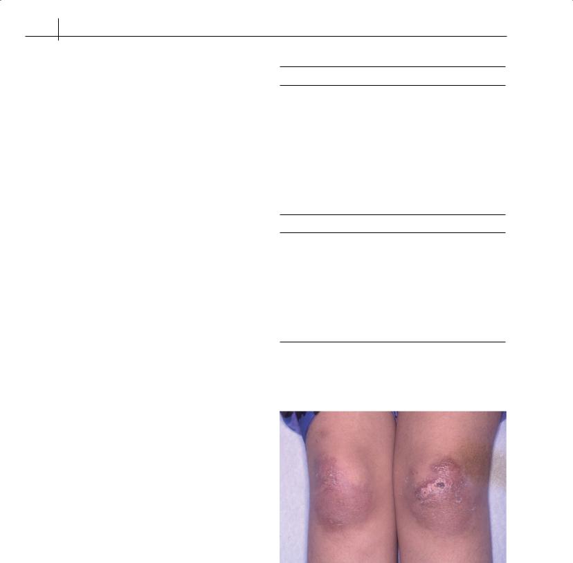
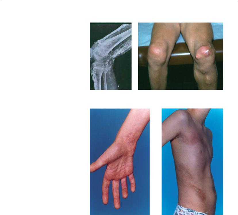
Juvenile dermatomyositis (JDM) may present at any age, with characteristic skin involvement (Figure 16.5), and proximal muscle weakness which can present acutely or indolently. JDM has a broad range of severity; contrary to adult-onset DM, there is no association with malignancy. Patients with JDM may develop calcinosis as a late complication of poorly controlled disease (Figure 16.6). Severe complications at the time of active disease include risk of aspiration pneumonia and interstitial lung disease. Diagnosis usually rests on clinical assessment, and elevated serum muscle enzymes; most children do not require electromyography or a muscle biopsy in the absence of atypical features. Magnetic resonance imaging of the muscles is very useful to demonstrate muscle involvement and monitor disease activity. Treatment of JDM requires rapid initiation of high-dose corticosteroids, frequently accompanied by methotrexate, although other medication such as intravenous immunoglobulin, cyclophosphamide or anti-cytokine agents are used in severe or refractory disease. Physical therapy input is essential to optimize outcome.
Table 16.3 SLE in children and adolescents
Common presenting symptoms
Malar rash
Arthritis
Fatigue
Fever
Weight loss
Oral ulcers
Alopecia
Pleuritis/pericarditis
Central nervous system findings
Photosensitivity
Raynaud’s phenomenon
Lymphadenopathy
Hepatosplenomegaly
Laboratory findings
Anaemia (may be haemolytic with positive red cell autoantibodies) Leukopaenia, lymphopaenia
Thrombocytopaenia Elevated liver enzymes
Elevated kidney function tests (blood urea nitrogen, creatinine) Decreased complement components C3 and C4
Positive antinuclear antibody
High titre positive anti-double-stranded DNA antibody
Positive autoantibodies to extractable antigens (anti-Ro (SSA); anti-La (SSB); anti-Sm; anti-RNP)
Positive antiphospholipid antibodies (anti-cardiolipin, lupus anticoagulant)
Figure 16.5 Gottron’s rash over the knees in juvenile dermatomyositis
MSK Disorders in Children and Adolescents |
103 |
|
|
|
|
Figure 16.6 Calcinosis in juvenile dermatomyositis
Figure 16.7 Localized scleroderma with morphoea skin lesions and underlying muscle wasting of the right hand
Sclerodermas
Scleroderma in childhood is rare and heterogeneous, and subtypes are determined by the type and number of lesions, the area of involvement and serological abnormalities. Localized scleroderma (Figure 16.7) is the most common and can present at any age, with the appearance of a patch of abnormal skin, which when untreated, generally follows a course of active expanding disease, fibrosis and eventual softening with some “remission”. The functional and cosmetic impact can be profound, as the lesions may interfere with
growth of a limb and subcutaneous tissues (of the face or a limb). Current practice advocates aggressive treatment regimes (corticosteroid and methotrexate) to control disease and limit severe disfigurement and disability. Systemic scleroderma is very rare in children and includes progressive diffuse fibrous changes of the skin and fibrous changes involving internal organs—most commonly lungs, gastrointestinal tract, heart and kidneys—with a significant mortality. Systemic scleroderma is slowly progressive, has a guarded prognosis and requires potent immunosuppression, although clinical trials are lacking to guide practice.
104 ABC of Rheumatology
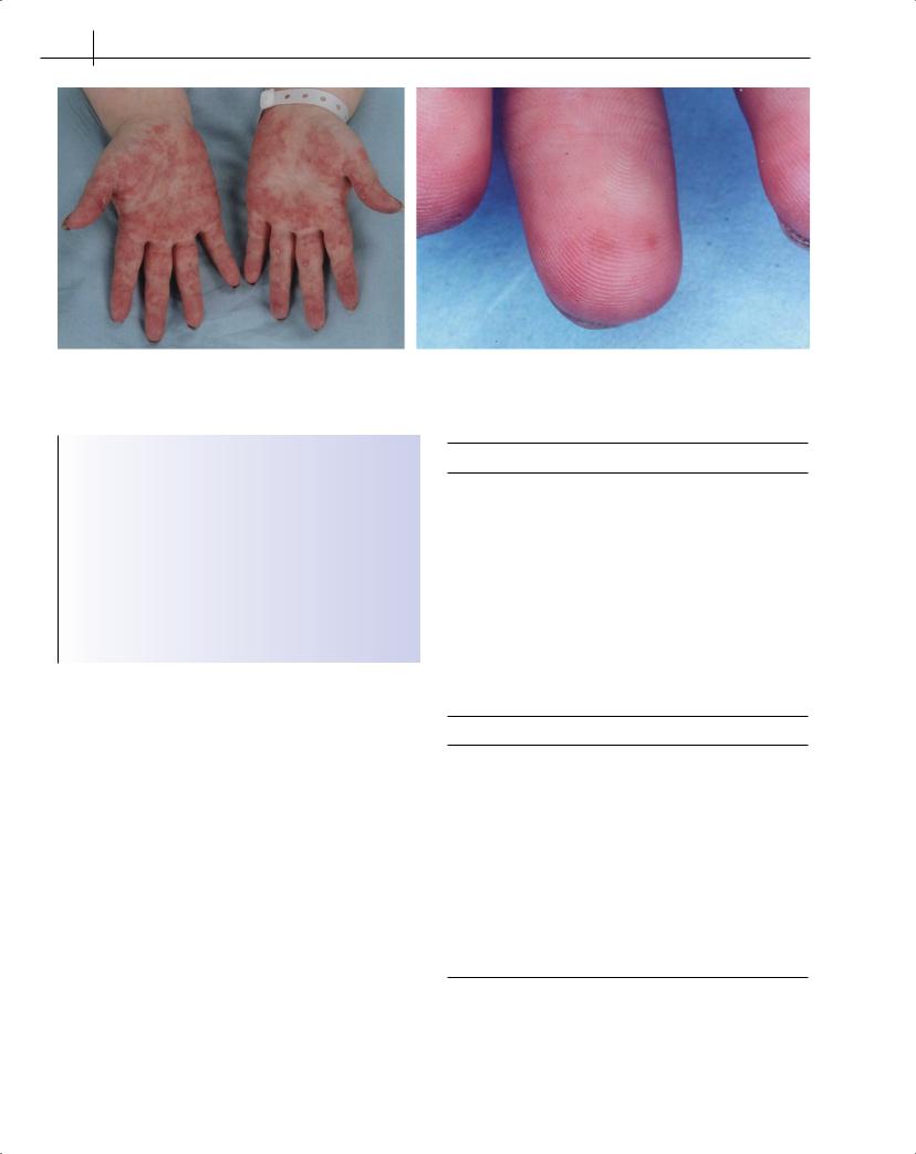
Figure 16.8 Vasculitis of the hands
Box 16.5 Features of Kawasaki disease
Fever for more than 5 days plus at least four of the following signs:
•Bilateral conjunctival injection
•Changes in the oropharyngeal membranes (swollen fissured lips, strawberry tongue, injected pharynx)
•Changes in peripheral extremities (erythema and swelling of hands and feet followed by desquamation of the skin)
•Polymorphous rash
•Cervical lymphadenopathy (at least one node >1.5 cm)
Vasculitis
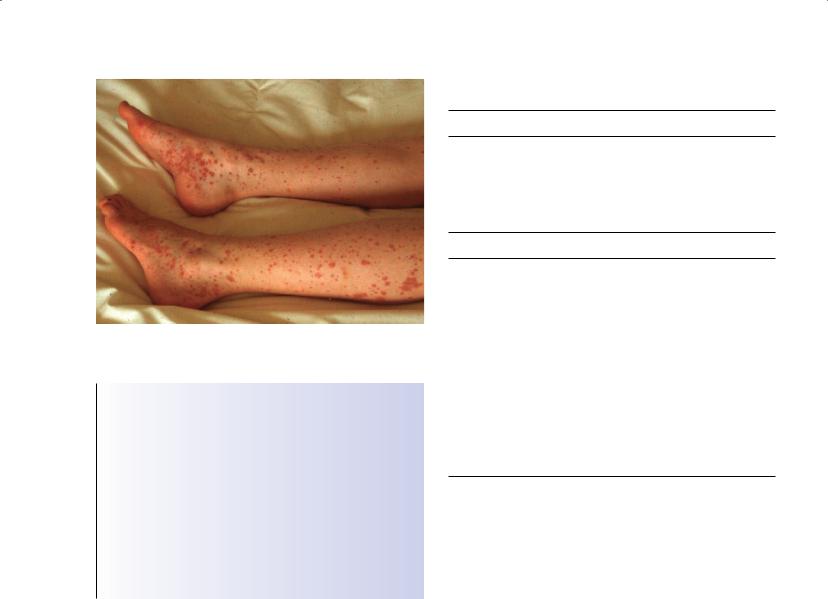
Vasculitis (Figure 16.8) is a heterogeneous group of disorders, most commonly classified by the size of involved blood vessels. A diagnosis may be suggested by multi-system clinical involvement and laboratory features (Table 16.4). The common childhood vasculitides Henoch–Schönlein purpura (HSP) and Kawasaki (KD) disease (Box 16.5) often have transient arthritis affecting large joints. HSP is characterized by palpable purpura (Figure 16.9) over the legs and buttocks, abdominal pain, haematuria and arthritis. In general, HSP resolves completely within 4 weeks of onset; however, some patients have recurrences of rash and gastrointestinal symptoms, and a small percentage of children who develop renal disease with HSP go on to renal failure. KD is an acute systemic vasculitis, predominantly in young children (less than 5 years); it is usually self-limiting but has the potential for causing severe long-term complications due to the involvement of coronary and other blood vessels with aneurysms. Prompt recognition of KD is essential in providing early treatment with intravenous immunoglobulin, which decreases the risk of developing coronary aneurysms significantly.
Table 16.4 Vasculitides in childhood
Type according to size of vessel
Large-vessel vasculitis
Takayasu’s arteritis
[Giant cell (temporal) arteritis—rarely seen in adolescents/children]
Medium-vessel vasculitis
Polyarteritis nodosa
Kawasaki disease
Small-vessel vasculitis
Wegener’s granulomatosis Churg–Strauss syndrome Microscopic polyangiitis Henoch–Schönlein purpura Cutaneous leukocytoclastic vasculitis
[Cryoglobulinaemic vasculitis—rarely seen in adolescents/children]
Features suggesting a vasculitis in adolescents and children
Clinical
Fever, weight loss, persistent fatigue
Skin rash: palpable purpura, vasculitic urticaria, nodules, ulcers Neurologic signs: headache, mononeuritis multiplex, focal CNS lesions Arthritis or arthralgia, myalgia or myositis
Hypertension
Pulmonary infiltrates or haemorrhage
Laboratory
Increased acute-phase reactants (ESR, CRP) Anaemia, leukocytosis
Eosinophilia
Antineutrophil cytoplasmic antibodies (ANCA)*
Elevated factor VIII-related antigen (von Willebrand factor) Haematuria
*ANCA: cytoplasmic (c-ANCA) associates specifically with Wegener’s granulomatosis and perinuclear (p-ANCA) associates with microscopic polyangiitis and a variety of other vasculitides
CRP = C-reactive protein; ESR = erythrocyte sedimentation rate
Reproduced by kind permission of the Arthritis Research Campaign (www. arc.org.UK)
MSK Disorders in Children and Adolescents |
105 |
|
|
|
|
Figure 16.9 Palpable purpura in Henoch–Schönlein purpura
Box 16.6 Inherited periodic fever syndromes
•Familial Mediterranean fever
•Hyerimmunoglobulinaemia D—also known as mevalonic kinase deficiency
•Tumor necrosis factor receptor-associated periodic syndrome
•The cryopyrinopathies:
Familial cold auto-inflammatory syndrome
Muckle–Wells syndrome
Chronic infantile neurologic cutaneous and articular syndrome—also known as neonatal-onset multi-system inflammatory disease
•Periodic fever with aphthous stomatitis, pharyngitis and adenitis
•Cyclic neutropenia
Table 16.5 Health-care transition planning in the paediatric rheumatology clinic
Planning for transition
Begin to discuss eventual transition from pediatrics with child and parent at an early stage (by puberty)
Engage the child in health-care visits directly at their developmental level
Begin to encourage the child/youth to spend time alone with health-care providers during visits
Transition tasks
Education about disease, prognosis, treatments and roles of health-care providers specifically directed at youth
Education about generic youth health issues (diet, dental care, sleep, exercise, smoking, alcohol, sexual health, recreational drugs)
Address adherence to medical plan at every visit
Career and vocational planning
Promote youth self-management, advocacy and independence in health-care decision-making
Assist with separation from parents with respect to medical issues
Acknowledge and be respectful of broader adolescent transitional tasks
Ultimately transfer them to adult rheumatologist and health-care providers (who are ideally youth-friendly) and forward adequate health records
Reproduced by kind permission of the Arthritis Research Campaign (www. arc.org.UK)
mandible can be involved. Radiographs show osteolytic changes similar to osteomyelitis, and a bone scan may show lesions that are asymptomatic. Antibiotics are not effective, and many children with CRMO have good symptomatic relief with non-steroidal antiinflammatory drugs. For those with persistent disease, bisphosphonates are often effective.
Rare inflammatory syndromes
Inherited auto-inflammatory syndromes
These syndromes (Box 16.6) are rare, and children may present with repeated unexplained bouts of fever with a variety of clinical findings associated with the fevers, which may include rash (Figure 16.1), MSK complaints, abdominal pain and ocular and neurologic complaints. There is often a broad range of severity among patients with the same genetic disorder, making diagnosis on clinical grounds often challenging. New treatment options have become available for some patients with periodic fever syndromes, and patients require specialist supervision.
Chronic recurrent multifocal osteomyelitis
Chronic recurrent multifocal osteomyelitis (CRMO) is a condition that presents similarly to bacterial osteomyelitis, but no organism can be isolated and there are often multiple involved sites with recurring episodes. Children or adolescents present with bone pain, sometimes accompanied by swelling; most common affected areas are long bones (tibia), but ribs, clavicle, vertebrae or
The role of the multidisciplinary team
The paediatric rheumatology multidisciplinary team (MDT) is highly skilled in the provision and coordination of comprehensive and often complex management regimes for the child and family, providing education and support, with other specialist services as well as community services, schools and health-care providers within shared care clinical networks. Many children with rheumatic diseases have continuing disease activity or relapses in adulthood, or sequelae from previous disease activity, which require ongoing medical treatment. Health-care transition, for youths with childhood-onset rheumatic diseases describes the movement of patients from childand family-centred paediatric care to adultoriented health-care systems. The MDT coordinate transitional care, addressing generic and disease-specific health issues (Table 16.5), and ultimately transfer to adult rheumatology services. Education and support are paramount, particularly with complex treatment regimes and the impact on adolescent behaviours, such as avoidance of pregnancy and excess alcohol in those taking methotrexate.

106 ABC of Rheumatology
References
Foster HE, Kay LJ, Friswell M, Coady DA, Myers A. pGALS—a paediatric musculoskeletal screening examination for school aged children based on the adult GALS screen. Arthritis Care Research 2006; 55: 709–716. A webstreamed demonstration of pGALS, supplementary materials and DVD are available online, at: http://www.arc.org.uk/arthinfo/emedia.asp
Malleson PN, Beauchamp P. Diagnosing musculoskeletal pain in children.
Canadian Medical Association Journal 2001; 165: 183–188.
Further reading
Cassidy JT, Petty RE. Textbook of Pediatric Rheumatology, 5th edn. WB Saunders, Philadelphia, PA, 2005.
Compeyrot-Lacassagne S, Feldman B. Inflammatory myopathies in children.
Pediatric Clinics of North America 2005; 52: 493–520.
Foster HE, Cabral DA. Is musculoskeletal history and examination so different in paediatrics? Best Practice & Research. Clinical Rheumatology 2006;
20: 241–262.
Klein-Gitelman, M, Reiff A, Silverman ED. Systemic lupus erythematosus in childhood. Rheumatic Disease Clinics of North America. 2002; 28: 561–577.
McDonagh JE. Transition of care from adult to paediatric rheumatology.
Archives of Disease in Childhood 2007; 97: 802–807.
Murray KJ, Laxer RM. Scleroderma in children and adolescents. Rheumatic Disease Clinics of North America 2002; 28: 603–624.
Ozen, S. The spectrum of vasculitis in children. Best Practice & Research Clinical Rheumatology 2002; 16: 411–425.