Материал: ТЭЛА

691062CPHXXX10.1177/1715163517691062C P J / R P CC P J / R P C research-article2017
PRACTICE TOOL  1&&3 3&7*&8&%
PRACTICE TOOL * PEER-REVIEWED
The management of venous thromboembolism: A practical tool for the front-line clinician
Tammy J. Bungard, BSP, PharmD; William Semchuk, BSP, PharmD, MSc
Case
You are just about to fnish your evening shif and close the pharmacy when Mr. James, a 57-year-old man who is well known to you, makes his way to the dispensary, favouring his right leg. He looks at you with a grimace on his face, and hands you 2 prescriptions, stating he just came from the local emergency department/ health care centre. You look at the prescriptions and see that one is for pain management and the other is an anticoagulant. You note the anticoagulant is for only 3 days. Mr. James says, “I have to go back and get some sort of test done on my leg during normal business hours tomorrow.”
Background
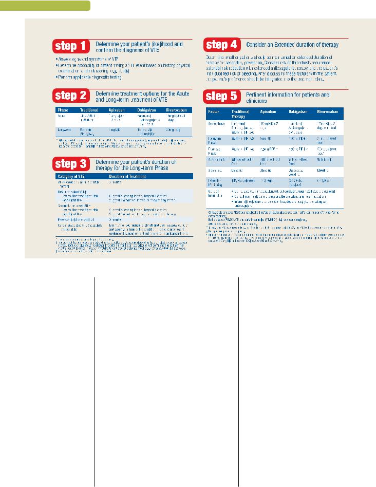
Venous thromboembolism (VTE), encompassing both deep vein thrombosis (DVT) and pulmonary embolism (PE), is common, afecting up to 5% of the population during their lifetime.1 Afer more than half a century with no new therapies, 3 novel oral anticoagulants (NOACs) are now approved by Health Canada for the treatment of acute VTE and prevention of recurrent VTE.2-4 Te NOACs ofer many advantages over the traditional therapy of a parenteral anticoagulant transitioned to warfarin, and as a result, there is an increasing shif to treating many of these patients outside of a hospital setting.5 Given this shif in care, front-line clinicians must be aware of nuances with these newer treatment options. Herein, we highlight a case to feature a practice tool in the form of a pocket card that provides a systematic, 5-step approach for front-line clinicians to aid in the management of patients across the care continuum of VTE
(Figure 1). Tis pocket card is available in full as Appendix 1 in the online version of the article and from the Canadian Cardiovascular Pharmacists Network at www.ccpn.ca.
Process for management of acute VTE
Step 1: Determine your patient’s likelihood of VTE and confrm diagnosis
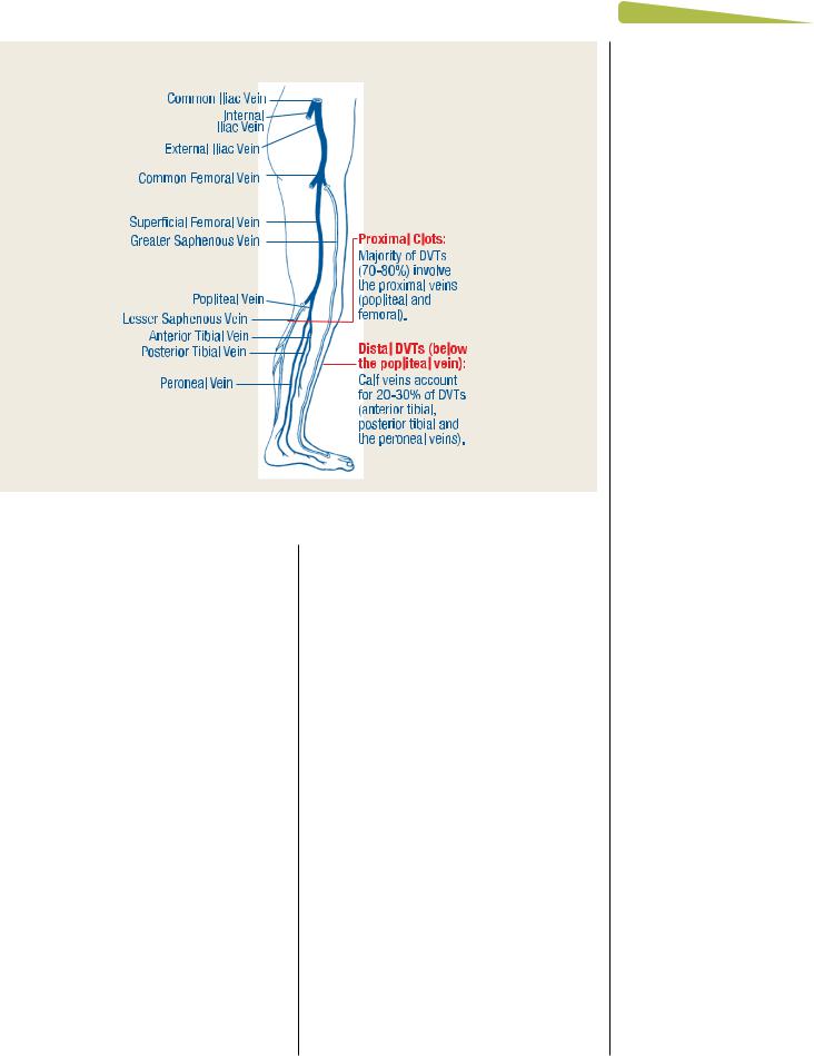
DVT occurs quickly and results in acute changes to the afected limb.6-8 A typical red, warm swollen unilateral limb occurs below the location of the clot given the lack of venous return and resultant increased pressure in the venous system with extravasation of fuid into the leg (see Appendix 1, Table 1). Clinically, diferentiation between proximal (above the knee, or clot location in the popliteal vein/more proximal) and distal DVTs is helpful, as proximal clots tend to be larger (occur in larger veins) and have an increased likelihood of embolization to the lungs (Figure 2).9 Te extent of Mr. James’s leg swelling is indicative of both the location and extensiveness (size) of the clot.10 While Mr. James’s presentation may be typical of an acute DVT, the nonspecifc presentation of DVT mandates objective diagnostic confrmation: Mr. James could also have cellulitis, a Baker’s cyst, lymphedema, hematoma, muscle tear, and so on. To proceed with testing for acute DVT, probability
scores may be used to discern the likelihood of a DVT (see Appendix 1, Table 2).11,12 Should
Mr. James have a low probability of DVT, progression with a D-dimer (a simple blood test for fbrin degradation products [evidence of
© The Author(s) 2017
DOI:10.1177/1715163517691062
$ 1 + 3 1 |
$ r M A R C H / A P R I L r 7 0 - / 0 |
8 1 |
13"$5*$& 500-
FIGURE 1 Steps in care (brief summary)
clot breakdown]) may be done.13 Te D-dimer is a very sensitive test with modest specifcity; therefore, it is most useful if it is negative (below threshold), which enables the clinician to rule out acute DVT (as there is no evidence of clot [fbrin] breakdown). Should it be positive, progression to compression ultrasound is required.
Depending on the geographic location, the ability to perform compression ultrasound may be limited to certain days of the week or hours of the day. Mr. James was sent home from his local health care centre with the presumption that he may have a DVT, with the need to return during business hours to rule the diagnosis in or out through ultrasound confrmation. Most patients who are clinically stable with acute DVT may be managed on an ambulatory basis; historically, many emergency rooms have administered a therapeutic dose of low-molecular-weight heparin (LMWH) and had the patient return daily for their LMWH injection until the diagnosis was ruled in or out.5 With the NOACs, an oral option is now available that has the ability to further simplify care and negate the need for injections
and costly emergency room visits as well as extensive laboratory monitoring. Notably, if Mr. James presented to his physician’s ofce with a moderate to high probability of DVT, it would be likely that progression to compression ultrasound would be done (performing a D-dimer is limited given the turnaround for community laboratories), with booking of an urgent ultrasound in the community and provision of anticoagulant therapy in the interim.
Mr. James has taken his anticoagulant and now his ultrasound is complete. He returns to the pharmacy with his report and hands it to you, and you read, “extensive clot in the popliteal vein extending into the superfcial femoral vein.” While the pharmacist is not diagnosing the clot, it is important for him or her to understand that the clot burden, along with the cause of the clot, may have implications on therapy duration and occurrence of complications. While you have your laboratory system open, you would look for other testing for Mr. James, specifcally renal function and complete blood count (CBC), as well as coagulation tests that may have been
8 2 |
$ 1 + 3 1 |
$ r M A R C H / A P R I L r 7 0 - / 0 |
13"$5*$& 500-
FIGURE 2 Lower extremity vasculature8
performed. Mr. James then proceeds to hand you another prescription and states, “Te doctor said the dose would change and that I’m to take this for at least 3 months.”
Step 2: Determine treatment options for the acute and long-term treatment of VTE
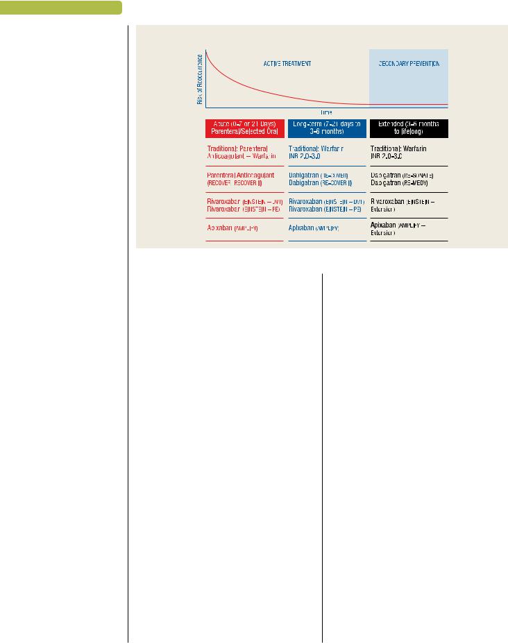
Tree phases of care have been described for the management of VTE (Figure 3).14 First, an acute phase (day 0 to as long as day 21) encompasses the initial occurrence/diagnosis of the clot, where clot propagation is likely to occur without therapy. Terapy in this phase is ofen with higher doses of oral agents or parenteral therapy (±warfarin) to treat the initial clot (see Appendix 1, Table 515-19 and Table 62-4). Second, a long-term active treatment phase (3 months to as long as 6 months) ensures the appropriate treatment of the initial clot with (typically) an oral anticoagulant agent (see Appendix 1, Table 6). Tird, an extended phase (also known as secondary prevention) may follow for those having a high risk of clot recurrence, balancing this risk with that of major bleeding. Tis later extended phase must be informed by patients’ values and preferences. For each of these phases, clinical trials have been performed using varying oral
anticoagulant regimens, as outlined in Figure 314 and Table 420-24 of Appendix 1.
Te goals of treatment (in the short term) for Mr. James are to use an anticoagulant to stabilize the existing clot to prevent clot extension or embolization and to allow his body to begin the process of breaking down the clot. Traditionally, Mr. James would have been limited to receiving only a parenteral anticoagulant (therapeutic doses of either LMWH or fondaparinux) with the intent of transitioning to warfarin therapy
(target international normalized ratio [INR] of 2.0-3.0; see Appendix 1, Table 5).25,26 While very
efective, use of this medication regimen on an ambulatory basis requires teaching patients to self-administer subcutaneous injections for a minimum of 5 days and obtainment of 2 consecutive days of therapeutic INRs (whichever is longer) with frequent laboratory testing. Several large-scale, noninferiority clinical trials have been conducted comparing the traditional standard of care (parenteral anticoagulant transitioned to warfarin) to each NOAC, and all have demonstrated that NOACs are noninferior to traditional therapy for the prevention of recurrent VTE and VTE-related death, with major bleeding rates similar or lower than that
$ 1 + 3 1 |
$ r M A R C H / A P R I L r 7 0 - / 0 |
8 3 |
13"$5*$& 500-
FIGURE 3 VTE phases of care and clinical trials14
reported with traditional care (see Appendix 1, Table 4).20-24
Mr. James presents as a stable patient with a proximal DVT and is typical of the population represented in the NOAC clinical tri- als.20-24 You recall reviewing his renal function (CrCl ~75 mL/min) and noted his hemoglobin within his CBC was within normal limits, providing a benchmark should he have a hemorrhagic event. Given this, treatment with any of the applicable NOAC regimens (see Appendix 1, Table 6) would be appropriate for Mr. James,2-4 and the regimens are recommended as a preferential choice over warfarin by the current Chest guidelines.27 It is important to note that clinical trials assessing the efcacy and safety of NOACs in the management of VTE fall into 2 general designs: apixaban and rivaroxaban studies have compared traditional therapy with an injectable anticoagulant transitioning to warfarin vs the NOAC monotherapy at a larger initiating dose moving to a maintenance dose (rivaroxaban, 15 mg twice daily [with food] must be adminis-
tered for 21 days, then stepping down to 20 mg daily with food21,22; apixaban, 10 mg twice daily
for 7 days, followed by 5 mg twice daily with no dosage adjustment based on weight, renal function and serum creatinine).20 In contrast, studies assessing dabigatran administered a parenteral
anticoagulant for 5 to 10 days prior to the transition to dabigatran 150 mg twice daily.23,24
For stroke prevention in atrial fbrillation, NOACs have varying dosage recommendations that are dependent on diferent variables for each agent, including renal dysfunction, advanced age (with or without accompanying bleeding risk factors) or low body weight. It is important to note that for the indication of acute VTE, provided the minimum renal function cutof is met, no dosage adjustment was made in the clinical trials. Clinicians should avoid rivaroxaban and dabigatran in patients with CrCl <30 mL/ min and apixaban if CrCl <25 mL/min, as these patients were excluded from the NOAC trials, and these agents accumulate in patients with signifcant renal dysfunction. Clinical trials also excluded patients having DVTs in atypical locations (e.g., splanchnic veins, hepatic veins, axillary veins, subclavian veins, etc.) or those having vena cava flters inserted. Moreover, within the population sufering a PE, those having massive clots requiring thrombolytic therapy or surgery or presenting with hemodynamic instability were excluded. Given the lack of data in these unique patient populations, most clinicians would elect to use traditional therapy at this time. Notably, a minority of patients having cancer were included in the NOAC trials. Current guidelines recommend LMWH therapy over oral anticoagulant options for cancer-associated thrombosis.27 Should an oral anticoagulant be used in cancerassociated thrombosis, no preference for either
8 4 |
$ 1 + 3 1 |
$ r M A R C H / A P R I L r 7 0 - / 0 |

warfarin or a NOAC is given.27 Te front-line clinician should be aware of characteristics of the NOACs, including (but not limited to) quick onset, quick ofset, extent of renal elimination, contraindications to use and drug-drug interactions (Table 1 and see Appendix 1, Table 828-30).
Step 3: Determine your patient’s duration of therapy for the long-term phase
One of the most clinically challenging areas in the management of VTE is establishing the duration of therapy. Guidelines suggest anticoagulating patients for a minimum of 3 months with reassessment to follow.27 Patients who have a provoking, strong risk factor (such as surgery) require the shortest duration of therapy, while those having unprovoked clots have longer therapy durations, given that clot recurrence is more likely in a setting where no causative factor can be identifed (and removed).27 Regardless of the clinical scenario, it is helpful to pose, at therapy initiation, an anticipated duration of therapy to avoid having patients continue on long-term therapy unnecessarily as well as to minimize the probability of nonadherence due to lack of knowledge of intended duration. In determining the duration of therapy for the “long-term phase,” your goal is to efectively treat the initial clot (see Appendix 1, Table 9). Reassessment of therapy at 3 months will typically reveal signs and symptoms of residual clot for patients having a more extensive clot burden or even complications from the initial clot. Te most common complication from DVT is postthrombotic syndrome, occurring in 25% to 50%,31 whereas for PE, complications include compromise of the right ventricle and chronic thromboembolic pulmonary hypertension (occurring in ~5%).27 Signs/symptoms that might suggest the need for extension of therapy for Mr. James’s DVT may include afected limb continuing to be larger, limb swelling progressing with limb use, a pale or dusky colour to the limb, reporting of limitations with using the limb and a dull ache to the limb with continued use (e.g., postthrombotic syndrome). For those with PE, reports of ongoing pain, shortness of breath and limitations in activity prior to the clot are common. Tese patients should be treated longer, with plans for ongoing reassessment (every 3-6 months). Patients having VTE in the setting of cancer should be treated with LMWH for a minimum of 3 to 6 months.
13"$5*$& 500-
Step 4: Consider an extended duration of therapy
Following the long-term treatment phase, the front-line clinician must, in conjunction with the patient, assess the need for ongoing therapy to prevent a recurrent VTE (i.e., extended or secondary prevention). Tis is one of the most clinically challenging areas in the management of VTE, given that we lack good prediction tools for the risk of recurrent VTE and risk of major bleeding and must strive to integrate patient values and preferences into the decisions made. Patients with transient provoking factors (e.g., surgery, hormone therapy) have the lowest risk of VTE recurrence and therefore would be expected to derive less beneft from extending therapy beyond 3 months (see Appendix 1, Table 10).27,32 In contrast, those having unprovoked VTEs have higher recurrence rates and thus may beneft more from extension therapy, provided the risk of major bleeding is not excessive and is minimized by removing factors that may predispose to bleeding (see Appendix 1, Table 11).27 Notably, factors identifed for the risk of major bleeding largely stem from data with traditional therapy (parenteral anticoagulant followed by warfarin), and given that we now have NOACs, these rates may be lower.
Several clinical trials have assessed NOACs
for the extended phase of therapy (see Appendix 1, Table 12).21,33,34 Most of these trials have
been placebo controlled, and the clinician must realize that clinical equipoise was evident for patients enrolled in these trials, given they had a 50% chance of receiving no ongoing therapy. In contrast, trials having a traditional care (namely, warfarin) control group would be expected to recruit patients for whom the clinician has made the decision that extended prevention is necessary and hence that recurrent VTE was more likely. All 3 placebo-controlled trials clearly showed efcacy for the NOAC to
prevent recurrent VTE, with no statistically signifcant increase in major bleeding.21,33,34
Notably, the only dose of dabigatran studied in VTE is the 150 mg twice-daily dosing.23,24,34
Despite this, the product monograph suggests applying the lower 110 mg twice-daily dose for those patients fulflling criteria for this dose for the indication of atrial fbrillation. 3 Te only NOAC having clinical trial data at a prophylactic dose for this extended phase is apixaban.33 Notably, both doses of apixaban (5 mg twice
$ 1 + 3 1 |
$ r M A R C H / A P R I L r 7 0 - / 0 |
8 5 |